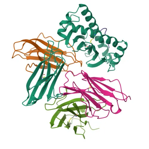
Monoclonal Antibodies
Rabbit anti-Human/Monkey CD8a Monoclonal Antibody for Flow Cytometry - P01732
Item Number : CM0008022
Price varies based on specs and customizations
- Application
- FC
- Cross Reactivity
- Human, Cynomolgus
- Protein Weight
- 21kDa/25kDa/30kDa

Shipping:
Contact us to get shipping details Enjoy On-time Dispatch Guarantee.
Why Choose Us
Easy ordering process, quality products, and dedicated support for your business success.
Core Product Specifications and Parameters
| Parameter | Value |
|---|---|
| Product Name | Rabbit anti-Human/Monkey CD8a mAb |
| Remarks/Alias | CD8; p32; Leu2; CD8alpha |
| Species | Human |
| GeneID (Human) | 925 |
| GeneID | 925 |
| Immunogen | Recombinant fusion protein containing a sequence corresponding to amino acids 22-182 of human CD8A (NP_001759.3) |
| Source | Rabbit |
| Category | Monoclonal Antibodies |
| Application | FC |
| Cross Reactivity | Human, Cynomolgus |
| SWISS | P01732 |
| Protein Weight | 21kDa/25kDa/30kDa |
| Shipping | Ice bag |
Biological Background: CD8a Function and Localization
- CD8A is T-cell surface glycoprotein CD8 alpha chain, also known as T8/Leu-2, encoded by the CD8A gene in humans. Alternative splicing produces multiple isoforms.
- Its core function is as an integral membrane glycoprotein coreceptor for MHC class I molecules. In T-cells, it interacts simultaneously with the T-cell receptor (TCR) and MHC class I:peptide complex, recruiting the Src kinase LCK to the TCR-CD3 complex to initiate intracellular signaling pathways leading to cytotoxic T-lymphocyte (CTL) activation, lymphokine production, and adhesion.
- CD8A homodimers on natural killer (NK) cells provide a survival mechanism, enabling conjugation and lysis of multiple target cells, and promote the differentiation of activated lymphocytes into memory CD8 T-cells.
- CD8A belongs to the immunoglobulin superfamily and typically forms disulfide-linked heterodimers with CD8B (CD8 beta chain) on thymus-derived T-cells; homodimers are also expressed on subsets of immune cells.
- Subcellular localization includes the cell membrane and secreted form. It is a single-pass type I membrane protein with a signal peptide, an extracellular immunoglobulin-like domain, a transmembrane helix, and a cytoplasmic tail.
- Tissue specificity: Expressed on cytotoxic T-cells, a subset of NK cells, memory T-cells, intraepithelial lymphocytes, monocytes, dendritic cells, and on plasmacytoid dendritic cells upon herpes simplex virus-1 stimulation.
- Post-translational modifications and keywords: Glycosylated, palmitoylated, and contains disulfide bonds. Annotated as a glycoprotein, lipoprotein, disulfide bond; involved in adaptive immunity, alternative splicing; linked to disease variants.
Experimental Guidance and Technical Tips
- The immunogen is a recombinant protein covering the extracellular region (aa 22-182). This design likely yields antibodies that recognize surface epitopes on live cells without permeabilization, making it well-suited for direct flow cytometry (FC) staining.
- For FC, titrate the conjugate to determine the optimal working dilution. Include appropriate isotype control antibodies and Fc receptor blocking. Verify specificity using known CD8A-positive cell lines (e.g., Jurkat) or peripheral blood mononuclear cells.
- Cross-reactivity is reported with cynomolgus monkey. If using cynomolgus samples, validate the antibody in your own assay system with proper controls.
- Multiple protein weights (21, 25, 30 kDa) are noted; these likely reflect differentially glycosylated forms or isoform variants. If performing western blot as a confirmatory step, expect potential multiple bands.
- For multicolor flow cytometry panels, confirm minimal spectral overlap with other fluorophores and include compensation controls. The rabbit monoclonal nature and direct conjugation facilitate reproducible results.
CamelBio: Your One-Stop Sourcing Bridge
CamelBio is a comprehensive IVD raw-material sourcing partner dedicated to aiding diagnostic manufacturers and research laboratories with validated biological reagents. This anti-CD8a monoclonal antibody, optimized for immune cell profiling, illustrates our ability to supply pre-conjugated flow cytometry antibodies, bulk ancillary reagents, and custom antigens. Whether you require validated antibody pairs, monoclonal/polyclonal antibodies for special targets, or rare target raw-material sourcing, CamelBio is committed to streamlining your development pipeline from concept to clinic.
Product Datasheet
Rabbit anti-Human/Monkey CD8a Monoclonal Antibody for Flow Cytometry - P01732
REQUEST A QUOTE
Our professional team will reply to you within one business day. Please feel free to contact us!