Monoclonal Antibodies
Rabbit anti-Human TLR1 Monoclonal Antibody for FC - Q15399
Item Number : CM0008446
Price varies based on specs and customizations
- Application
- FC
- Cross Reactivity
- Human
- Protein Weight
- 90kDa

Shipping:
Contact us to get shipping details Enjoy On-time Dispatch Guarantee.
Why Choose Us
Easy ordering process, quality products, and dedicated support for your business success.
Core Product Specifications and Parameters
| Parameter | Value |
|---|---|
| Product Name | Rabbit anti-Human TLR1 mAb |
| Remarks/Alias | TIL; CD281; rsc786; TIL. LPRS5 |
| Species | Human |
| GeneID (Human) | 7096 |
| GeneID | 7096 |
| Immunogen | Synthetic peptide (a synthetic peptide corresponding to a sequence within amino acids 325-424 of human TLR1 (NP_003254.2)) |
| Source | Rabbit |
| Category | Monoclonal Antibodies |
| Application | FC |
| Cross Reactivity | Human |
| SWISS | Q15399 |
| Protein Weight | 90kDa |
| Shipping | Ice bag |
Biological Background: TLR1 Function and Localization
- Toll-like receptor 1 (TLR1), also known as TIL or CD281, is a member of the Toll-like receptor family, which plays a fundamental role in pathogen recognition and activation of innate immunity.
- TLR1 specifically recognizes diacylated and triacylated lipopeptides from bacteria, and cooperates with TLR2 to mediate the innate immune response to bacterial lipoproteins. Related references: PMID:21078852
- Upon ligand binding, TLR1 forms an activation cluster with TLR2 and CD14 on the cell membrane, which triggers signaling via the MYD88-TRAF6 pathway, leading to NF-kappa-B activation, inflammatory cytokine secretion, and subsequent trafficking to the Golgi in a lipid-raft dependent manner. Related references: PMID:16880211
- TLR1 is localized at the cell membrane, membrane rafts, cytoplasmic vesicles, phagosome membrane, and the Golgi apparatus, reflecting its dynamic trafficking upon activation.
- Ubiquitously expressed, with highest levels in spleen, ovary, peripheral blood leukocytes, thymus, and small intestine, TLR1 plays a broad role in immune surveillance.
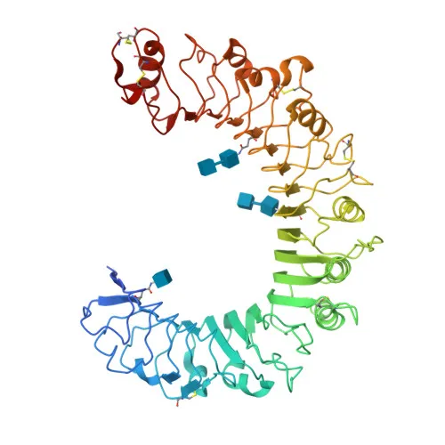
- Post-translational modifications include glycosylation and disulfide bonds, and the protein contains leucine-rich repeats typical of pattern recognition receptors.
Experimental Guidance and Technical Tips
- The antibody was raised against a synthetic peptide corresponding to amino acids 325-424 within the extracellular domain of human TLR1; this region is predicted to be involved in ligand recognition and may be accessible for flow cytometry detection.
- As this antibody is directly conjugated to , it is optimized for flow cytometric analysis (FC). We recommend titrating the antibody to determine the optimal concentration for your specific cell type and experimental conditions.
- Use appropriate isotype controls and consider co-staining with markers for immune cell subpopulations (e.g., CD14, TLR2) to assess TLR1 expression on monocytes, dendritic cells, or other leukocytes.
- Note that TLR1 can exist as a heterodimer with TLR2; thus, activation state may influence detection. Validate in your sample system and consider fixation/permeabilization protocols if detecting intracellular pools.
CamelBio: Your One-Stop Sourcing Bridge
CamelBio supports diagnostic manufacturers and research laboratories with a comprehensive range of IVD raw materials, including validated antibody pairs and monoclonal antibodies for innate immunity targets such as TLR1. Leveraging a global supply network, we offer bulk ancillary reagents and one-stop sourcing solutions to accelerate your assay development, from concept to clinic. Whether you need specialized antibody conjugates, rare target reagents, or technical consulting, CamelBio is your trusted partner in immunology research and IVD development.
REQUEST A QUOTE
Our professional team will reply to you within one business day. Please feel free to contact us!