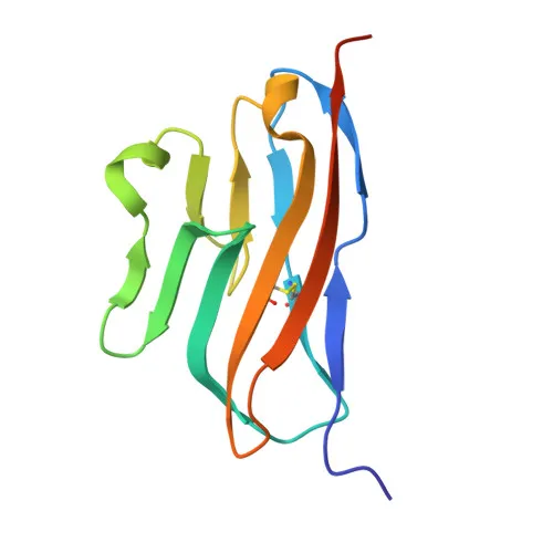
Monoclonal Antibodies
Rabbit anti-Human TIGIT Monoclonal Antibody for Flow Cytometry - Q495A1
Item Number : CM0010639
Price varies based on specs and customizations
- Application
- FC
- Cross Reactivity
- Human
- Protein Weight
- 18kDa/26kDa

Shipping:
Contact us to get shipping details Enjoy On-time Dispatch Guarantee.
Why Choose Us
Easy ordering process, quality products, and dedicated support for your business success.
Core Product Specifications and Parameters
| Parameter | Value |
|---|---|
| Product Name | Rabbit anti-Human TIGIT mAb |
| Remarks/Alias | VSIG9; VSTM3; WUCAM |
| Species | Human |
| Gene ID (Human) | 201633 |
| Immunogen | Recombinant protein or recombinant fusion protein containing a sequence corresponding to amino acids 22-141 of human TIGIT (NP_776160.2). |
| Source | Rabbit |
| Category | Monoclonal Antibodies |
| Application | FC |
| Cross Reactivity | Human |
| SWISS | Q495A1 |
| Protein Weight | 18kDa/26kDa |
| Shipping | Ice bag |
Biological Background: TIGIT Function and Localization
- TIGIT (T-cell immunoreceptor with Ig and ITIM domains), also known as VSIG9, VSTM3, or WUCAM, is an inhibitory receptor of the immunoglobulin superfamily.
- It suppresses T-cell activation by promoting the generation of mature immunoregulatory dendritic cells. Related references: PMID:19011627
- Upon binding to PVR/CD155 or NECTIN2/CD112 on antigen-presenting cells, TIGIT delivers inhibitory signals by recruiting Src kinases, GRB2, and SHIP1 to inhibit PI3K/MAPK pathways, and associates with beta-arrestin-2 to suppress NF-kappa-B signaling via TRAF6. Related references: PMID:23154388 PMID:24817116
- TIGIT also functions as a receptor for NECTIN4, inhibiting NK cell cytotoxicity. Related references: PMID:32503945
- It is localized at the cell membrane, consistent with its role in immune synapse modulation.
- TIGIT is expressed at low levels on peripheral memory and regulatory CD4+ T-cells and NK cells, and is upregulated following activation.
- Post-translational modifications include glycosylation and phosphorylation; the protein contains a disulfide bond and an ITIM domain.
Experimental Guidance and Technical Tips
- This -conjugated antibody is designed for direct flow cytometry; use appropriate isotype controls and optimize titration for your specific cell type and activation state.
- The immunogen corresponds to the extracellular region (aa 22-141), ensuring recognition of surface-expressed TIGIT on live cells.
- Since TIGIT is upregulated upon T-cell and NK cell activation, consider stimulating cells (e.g., with anti-CD3/CD28) prior to staining to enhance detection.
- For multicolor flow cytometry panels, verify compensation settings with single-stained controls and include viability dyes to exclude dead cells.
- Confirm the molecular weight band pattern (~18 kDa core, ~26 kDa glycosylated) if performing complementary western blot validation.
CamelBio: Your One-Stop Sourcing Bridge
CamelBio provides a one-stop sourcing bridge for IVD raw materials, including this high-quality anti-TIGIT monoclonal antibody for immune checkpoint research and diagnostic assay development. Our comprehensive portfolio includes validated antibody pairs, optimized monoclonal/polyclonal antibodies, bulk ancillary reagents, and rare target raw-material sourcing. Let CamelBio accelerate your project from concept to clinic with reliable supply and expert support.
Product Datasheet
Rabbit anti-Human TIGIT Monoclonal Antibody for Flow Cytometry - Q495A1
REQUEST A QUOTE
Our professional team will reply to you within one business day. Please feel free to contact us!