Monoclonal Antibodies
Rabbit anti-Human Syk Monoclonal Antibody for Intracellular Flow Cytometry - P43405
Item Number : CM0011130
Price varies based on specs and customizations
- Application
- FC (intra)
- Cross Reactivity
- Human
- Protein Weight
- 72kDa/70kDa

Shipping:
Contact us to get shipping details Enjoy On-time Dispatch Guarantee.
Why Choose Us
Easy ordering process, quality products, and dedicated support for your business success.
Core Product Specifications and Parameters
| Parameter | Value |
|---|---|
| Product Name | Rabbit anti-Human Syk mAb |
| Remarks/Alias | IMD82; p72-Syk |
| Species | Human |
| GeneID (Human) | 6850 |
| GeneID | 6850 |
| Immunogen | Synthetic peptide|A synthetic peptide corresponding to a sequence within amino acids 251-350 of human Syk (NP_003168.2) |
| Source | Rabbit |
| Category | Monoclonal Antibodies |
| Application | FC (intra) |
| Cross Reactivity | Human |
| SWISS | P43405 |
| Protein Weight | 72kDa/70kDa |
| Shipping | Ice bag |
Biological Background: Syk Function and Localization
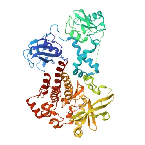
- Syk (Spleen Tyrosine Kinase) is a 72 kDa non-receptor tyrosine kinase characterized by two N-terminal SH2 domains and a C-terminal kinase domain, belonging to the SYK/ZAP70 family.
- It mediates signal transduction downstream of immune receptors including the B-cell receptor (BCR) and T-cell receptor (TCR), phosphorylating key adaptors like BLNK and effectors such as PLCG1 (PubMed:12456653, PubMed:15388330, PubMed:34634301). Related references: PMID:12456653 PMID:15388330 PMID:34634301
- SYK is essential for B-cell maturation at the pro-B to pre-B transition and is widely expressed in hematopoietic cells, including B-cells and neutrophils (PubMed:12456653, PubMed:8163536). Related references: PMID:12456653 PMID:8163536
- In innate immunity, SYK is activated by CLEC7A in response to fungi, promoting ROS production and inflammasome activation; it is required for IL15-stimulated neutrophil phagocytosis (PubMed:15123770). Related references: PMID:15123770
- SYK localizes to the cell membrane and cytoplasm, assembling into signaling complexes with activated receptors.
- Its activity is regulated by phosphorylation on multiple tyrosine residues upon receptor engagement, functioning as a central phospho-signaling hub.
- Beyond immunity, SYK participates in integrin-mediated cell adhesion (PubMed:12387735), osteoclast maturation, platelet activation, and vascular development. Related references: PMID:12387735
Experimental Guidance and Technical Tips
- The immunogen peptide maps to amino acids 251‑350 (within the kinase domain) of human Syk. The antibody may recognize full‑length Syk and splice variants containing this region.
- For intracellular flow cytometry, fix and permeabilize cells according to validated protocols. Include an appropriate isotype control and titrate the antibody for optimal staining.
- Validate staining on known Syk‑expressing cell models (e.g., Ramos B‑cell line, Jurkat T‑cell line, or human PBMCs).
- is a bright, photostable far‑red conjugate. For multicolor panels, perform careful compensation and ensure there is minimal spectral overlap with other red/far‑red fluorophores.
CamelBio: Your One-Stop Sourcing Bridge
CamelBio provides diagnostic manufacturers and research laboratories with one‑stop access to high‑quality IVD raw materials and expert technical support, covering every stage from concept to clinic. This anti‑Syk monoclonal antibody supports research into immune signaling pathways and can be integrated into diagnostic workflows for immune function assessment. CamelBio also offers validated antibody pairs, bulk ancillary reagents, and customized sourcing for rare targets to accelerate your IVD development.
REQUEST A QUOTE
Our professional team will reply to you within one business day. Please feel free to contact us!