Monoclonal Antibodies
Rabbit anti-Human PTH1R Monoclonal Antibody () for Flow Cytometry - Q03431
Item Number : CM0010046
Price varies based on specs and customizations
- Application
- FC
- Cross Reactivity
- Human
- Protein Weight
- 66kDa

Shipping:
Contact us to get shipping details Enjoy On-time Dispatch Guarantee.
Why Choose Us
Easy ordering process, quality products, and dedicated support for your business success.
Easy Process
Quality Assured
Dedicated Support
Core Product Specifications and Parameters
| Parameter | Value |
|---|---|
| Product Name | Rabbit anti-Human PTH1R mAb |
| Remarks/Alias | PFE; EKNS; PTHR; PTHR1 |
| Species | Human |
| Gene ID | 5745 |
| Immunogen | Recombinant fusion protein containing a sequence corresponding to amino acids 27-188 of human PTH1R (NP_000307.1) |
| Source | Rabbit |
| Category | Monoclonal Antibodies |
| Application | FC |
| Cross Reactivity | Human |
| SWISS | Q03431 |
| Protein Weight | 66kDa |
| Shipping | Ice bag |
Biological Background: PTH1R Function and Localization
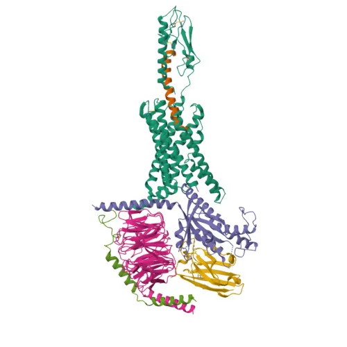
- PTH1R is a G protein-coupled receptor that binds parathyroid hormone (PTH) and PTH-related peptide (PTHLH), initiating intracellular signaling cascades (PubMed:10913300, 18375760, 19674967). Related references: PMID:10913300 PMID:18375760 PMID:19674967
- It couples to G(s) alpha proteins to activate adenylate cyclase, leading to increased cAMP levels (PubMed:20172855, 30975883). Related references: PMID:20172855 PMID:30975883
- PTHLH dissociates from PTH1R more rapidly than PTH, causing a faster decay of the cAMP response (PubMed:35932760). Related references: PMID:35932760
- The receptor is located at the cell membrane.
- Expressed in most tissues, with highest levels in kidney, bone, and liver.
- Contains key structural features including disulfide bonds, glycosylation sites, and phosphorylation sites; associated with diseases such as dwarfism.
Experimental Guidance and Technical Tips
- The immunogen corresponds to the N-terminal extracellular domain (amino acids 27-188) of human PTH1R, which is accessible on the cell surface without permeabilization. This design supports detection in live-cell flow cytometry.
- For flow cytometry, consider titrating the -conjugated antibody to determine the optimal staining dilution (e.g., 1:50 – 1:200) using cells known to express PTH1R, such as kidney or bone cell lines.
- Use appropriate negative controls (e.g., isotype control or cells with low/absent PTH1R expression) to set gates and assess specificity.
- The large Stokes shift of minimizes spectral overlap; verify instrument compatibility (excitation ~650 nm, emission ~665 nm).
- Fixation with aldehydes may alter the epitope; if fixation is required, test whether it affects antibody binding.
CamelBio: Your One-Stop Sourcing Bridge
- CamelBio is a trusted one-stop partner for IVD raw-material procurement, offering a full spectrum of validated antibody pairs, optimized monoclonal and polyclonal antibodies, bulk ancillary reagents, and custom sourcing solutions for rare targets.
- This anti-PTH1R antibody, targeting the parathyroid hormone receptor, supports research and diagnostic development in endocrinology, bone metabolism, and calcium homeostasis disorders.
- From early concept through clinical application, CamelBio bridges your sourcing gaps with reliable quality and expert technical support.
REQUEST A QUOTE
Our professional team will reply to you within one business day. Please feel free to contact us!