Monoclonal Antibodies
Rabbit anti-Human P-cadherin Monoclonal Antibody for Flow Cytometry - P22223
Item Number : CM0009580
Price varies based on specs and customizations
- Application
- FC
- Cross Reactivity
- Human
- Protein Weight
- 87kDa/91kDa

Shipping:
Contact us to get shipping details Enjoy On-time Dispatch Guarantee.
Why Choose Us
Easy ordering process, quality products, and dedicated support for your business success.
Core Product Specifications and Parameters
| Parameter | Value |
|---|---|
| Product Name | Rabbit anti-Human P-cadherin mAb |
| Remarks/Alias | CDHP; HJMD; PCAD |
| Species | Human |
| GeneID | 1001 |
| Immunogen | Recombinant protein: Recombinant fusion protein containing a sequence corresponding to amino acids 108-654 of human P-cadherin (NP_001784.2) |
| Source | Rabbit |
| Category | Monoclonal Antibodies |
| Application | FC |
| Cross Reactivity | Human |
| SWISS | P22223 |
| Protein Weight | 87kDa/91kDa |
| Shipping | Ice bag |
Biological Background: P-cadherin Function and Localization
- Cadherin-3, also known as Placental cadherin (P-cadherin), is a classical cadherin that mediates calcium-dependent homophilic cell-cell adhesion, playing a key role in tissue morphogenesis and maintenance.
- The CDH3 gene (GeneID: 1001) encodes multiple isoforms; the canonical isoform has a calculated molecular mass of ~91.4 kDa, consistent with observed bands at 87–91 kDa due to glycosylation.
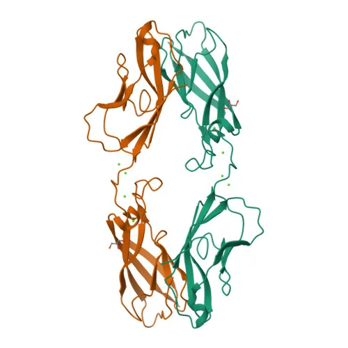
- P-cadherin is a single-pass type I transmembrane protein localized predominantly at the cell membrane, with extracellular cadherin repeats that bind calcium ions to stabilize adhesive junctions.
- Expression is detected in specific normal epithelial tissues (such as basal layers of stratified epithelia) and is upregulated in certain carcinoma cell lines, linking it to tumor biology and epithelial-mesenchymal transition.
- Functional keywords include cell adhesion, calcium binding, and sensory transduction/vision; disease-associated mutations in CDH3 cause ectodermal dysplasia, ectrodactyly, macular dystrophy (EEM syndrome) and hypotrichosis with juvenile macular dystrophy (HJMD).
- Post-translational modifications such as cleavage at dibasic sites (e.g., pro-peptide processing) and N-linked glycosylation are critical for cadherin maturation and adhesive function.
Experimental Guidance and Technical Tips
- Since this antibody is designed for flow cytometry, it is directly conjugated to , minimizing secondary detection steps; titrate the antibody (e.g., 0.1–1 µg per 10^6 cells) to optimise signal-to-noise in your experimental system.
- The immunogen comprises residues 108–654 of the extracellular domain, which is expected to generate antibodies against the mature, surface-exposed form of P-cadherin; confirm specificity in your model by assessing staining of known positive (e.g., A431) and negative cell lines.
- Include appropriate controls such as isotype-matched rabbit IgG-and fluorescence-minus-one (FMO) controls to validate FC gating strategies.
- Because P-cadherin is subject to cleavage, consider brief activation of cells (e.g., with trypsin/EDTA) and note that surface expression may vary with cell density and calcium concentration.
CamelBio: Your One-Stop Sourcing Bridge
CamelBio is your one-stop sourcing partner for IVD raw materials, offering a comprehensive range of validated antibody pairs, recombinant proteins, and ancillary reagents. For P-cadherin research and diagnostic development, we provide high-quality monoclonal antibodies like this conjugate, along with bulk ordering options and custom development services to accelerate your assay pipeline.
Product Datasheet
Rabbit anti-Human P-cadherin Monoclonal Antibody for Flow Cytometry - P22223
REQUEST A QUOTE
Our professional team will reply to you within one business day. Please feel free to contact us!