Monoclonal Antibodies
Rabbit anti-Human P-cadherin mAb - P22223
Item Number : CM0010429
Price varies based on specs and customizations
- Application
- FC
- Cross Reactivity
- Human
- Protein Weight
- 87kDa/91kDa

Shipping:
Contact us to get shipping details Enjoy On-time Dispatch Guarantee.
Why Choose Us
Easy ordering process, quality products, and dedicated support for your business success.
Core Product Specifications and Parameters
| Parameter | Value |
|---|---|
| Product Name | Rabbit anti-Human P-cadherin mAb |
| Remarks/Alias | CDHP; HJMD; PCAD |
| Species | Human |
| Gene ID | 1001 |
| Immunogen | Recombinant fusion protein containing a sequence corresponding to amino acids 108-654 of human P-cadherin (NP_001784.2). |
| Source | Rabbit |
| Category | Monoclonal Antibodies |
| Application | FC |
| Cross Reactivity | Human |
| SWISS | P22223 |
| Protein Weight | 87kDa/91kDa |
| Shipping | Ice bag |
Biological Background: Cadherin-3 Function and Localization
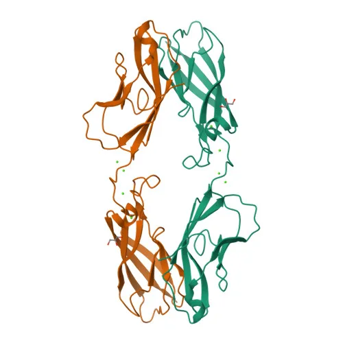
- Cadherin-3, also known as placental cadherin (P-cadherin), is a calcium-dependent cell adhesion protein encoded by the CDH3 gene.
- It mediates homophilic cell–cell interactions, preferentially binding to identical cadherin molecules on adjacent cells, contributing to the sorting of heterogeneous cell types.
- The protein is a single-pass type I membrane glycoprotein and member of the cadherin superfamily.
- Subcellular localization is predominantly at the cell membrane.
- Expression is observed in certain normal epithelial tissues and is also found in some carcinoma cell lines, implicating a role in carcinogenesis.
- Alternative splicing produces multiple isoforms; the reported molecular weights include 87 kDa and 91 kDa.
- Disease-associated mutations in CDH3 are linked to ectodermal dysplasia and hypotrichosis with juvenile macular dystrophy (HJMD), indicating roles in skin and hair follicle integrity and vision.
- The protein undergoes post-translational modifications including proteolytic cleavage and glycosylation, which may regulate its function.
Experimental Guidance and Technical Tips
- As a fluorophore-conjugated monoclonal antibody, it is designed for direct flow cytometry; start with a titration of 0.5–5 µg per 10⁶ cells and optimize for your specific cell type.
- Validate staining specificity using a relevant isotype control and confirm expression in known P-cadherin-positive human epithelial/carcinoma cell lines.
- Since the immunogen encompasses the extracellular domain (aa 108–654), the antibody likely binds an epitope accessible on live, non-permeabilized cells—compatible with surface staining protocols.
- Given the reported molecular weight variations (87/91 kDa), potential isoforms or glycosylation states may affect migration; validate detection in your sample system.
CamelBio: Your One-Stop Sourcing Bridge
CamelBio supports diagnostic developers and research labs with comprehensive access to IVD raw materials for cell adhesion and oncology targets. We offer validated antibody pairs, optimized monoclonal antibodies like this anti-P-cadherin clone, and bulk ancillary reagents to streamline your assay development. For rare or custom targets, our sourcing network bridges the gap from concept to clinic.
REQUEST A QUOTE
Our professional team will reply to you within one business day. Please feel free to contact us!