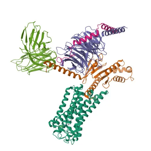
Monoclonal Antibodies
Rabbit anti-Human OPN1LW/OPN1MW mAb - P04000 / P04001
Item Number : CM0011623
Price varies based on specs and customizations
- Application
- FC
- Cross Reactivity
- Human
- Protein Weight
- 40kDa

Shipping:
Contact us to get shipping details Enjoy On-time Dispatch Guarantee.
Why Choose Us
Easy ordering process, quality products, and dedicated support for your business success.
Core Product Specifications and Parameters
| Parameter | Value |
|---|---|
| Product Name | Rabbit anti-Human OPN1LW/OPN1MW mAb |
| Remarks/Alias | CBP; RCP; ROP; CBBM; COD5; CBD; GCP; GOP; OPN1MW1 |
| Species | Human |
| GeneID (Human) | 5956/2652 |
| GeneID | 5956/2652 |
| Immunogen | Recombinant fusion protein containing a sequence corresponding to amino acids 1-48 of human OPN1LW/OPN1MW (NP_064445.2) |
| Source | Rabbit |
| Category | Monoclonal Antibodies |
| Application | FC |
| Cross Reactivity | Human |
| SWISS | P04000/P04001 |
| Protein Weight | 40kDa |
| Shipping | Ice bag |
Biological Background: OPN1LW/OPN1MW Function and Localization
- OPN1LW encodes long-wave-sensitive opsin 1, also known as red cone photoreceptor pigment or red-sensitive opsin (ROP).
- It functions as a G protein-coupled photoreceptor that selectively activates G(i) proteins upon exposure to long-wavelength (red) light, leading to a decrease in intracellular cAMP levels. Related references: PMID:2937147, PMID:6140680
- Photoactivation triggers isomerization of bound cis-retinal to all-trans-retinal, inducing a conformational change that initiates the G protein-mediated phototransduction cascade.
- OPN1LW is critical for mediating visual perception of red light in cone photoreceptor cells. Related references: PMID:12051694, PMID:8213841, PMID:8666378
- It localizes to the membrane of photoreceptor outer segments, specifically in cone cell outer segments.
- Expressed in red cone cells, alongside OPN1MW (green) and OPN1SW (blue) opsins, enabling trichromatic color vision.
- The protein is glycosylated, phosphorylated, and contains conserved disulfide bonds; it is classified under disease variants and belongs to the GPCR family involved in sensory transduction.
Experimental Guidance and Technical Tips
- The immunogen (N-terminal amino acids 1-48) is shared between OPN1LW and OPN1MW, suggesting this antibody may recognize both opsins; users should verify specificity in their experimental context.
- For flow cytometry (FC), optimize antibody dilution and staining protocols using appropriate positive and negative control cell lines expressing red/green cone opsins.
- Since OPN1LW/OPN1MW are membrane proteins, surface staining on live, unfixed cells may be feasible; however, fixation and permeabilization conditions should be empirically determined.
- Confirm that the fluorophore detection channel is properly compensated and free from spectral overlap in multicolor panels.
- Cross-reactivity is reported for human; reactivity with other species should be validated separately.
CamelBio: Your One-Stop Sourcing Bridge
CamelBio provides diagnostic manufacturers and researchers with streamlined access to high-quality IVD raw materials, including validated antibodies for vision and photoreceptor targets. Beyond this anti-OPN1LW/OPN1MW monoclonal antibody, we supply antibody pairs, optimized recombinant proteins, and ancillary reagents to support your assay development. Our sourcing expertise also covers rare targets and custom raw-material procurement, helping you move from concept to clinic with confidence.
REQUEST A QUOTE
Our professional team will reply to you within one business day. Please feel free to contact us!