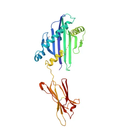
Monoclonal Antibodies
Rabbit anti-Human MICA/MICB Monoclonal Antibody for Flow Cytometry - Q29983 / Q29980
Item Number : CM0012053
Price varies based on specs and customizations
- Application
- FC
- Cross Reactivity
- Human
- Protein Weight
- 4kDa/31kDa/37kDa/43kDa

Shipping:
Contact us to get shipping details Enjoy On-time Dispatch Guarantee.
Why Choose Us
Easy ordering process, quality products, and dedicated support for your business success.
Core Product Specifications and Parameters
| Parameter | Value |
|---|---|
| Product Name | Rabbit anti-Human MICA/MICB mAb |
| Remarks/Alias | MIC-A; PERB11.1; PERB11.2; MICB |
| Species | Human |
| GeneID (Human) | 100507436/4277 |
| GeneID | 100507436/4277 |
| Immunogen | Recombinant fusion protein containing a sequence corresponding to amino acids 23-298 of human MICB (NP_005922.2). |
| Source | Rabbit |
| Category | Monoclonal Antibodies |
| Application | FC |
| Cross Reactivity | Human |
| SWISS | Q29983/Q29980 |
| Protein Weight | 4kDa/31kDa/37kDa/43kDa |
| Shipping | Ice bag |
Biological Background: MICA Function and Localization
- MICA functions as a stress-induced ligand for the activating receptor NKG2D, which is expressed on essentially all human natural killer (NK) cells, γδ T cells, and CD8+ αβ T cells. Related references: PMID:11491531, PMID:11777960
- Engagement of NKG2D by MICA triggers NK cell-mediated cytotoxicity and cytokine production, playing a critical role in anti-tumor and anti-viral immune responses. Related references: PMID:10426993
- MICA expression is upregulated under conditions of cellular stress, such as viral or bacterial infections and DNA damage, serving as a danger signal to eliminate compromised cells.
- Subcellularly, MICA is predominantly localized to the cell membrane, with some cytoplasmic presence.
- Widely expressed in tissues except the central nervous system; particularly abundant in gastric epithelium, keratinocytes, endothelial cells, and various epithelial tumors.
- Post-translational modifications include glycosylation, disulfide bond formation, and palmitoylation; it is also a target for ubiquitination (as inferred from UniProt keywords).
Experimental Guidance and Technical Tips
- This antibody is directly conjugated to , a bright and photostable fluorophore optimized for flow cytometry.
- For optimal staining, titrate the antibody using relevant human cell lines or primary cells known to express MICA/MICB under normal or stress conditions.
- Include appropriate isotype controls and consider Fc receptor blocking to reduce non-specific binding.
- Validate antibody performance in human PBMCs, tumor cell lines, or stressed epithelial cells to confirm detection of surface MICA/MICB.
- If using for intracellular staining, verify that the fixation/permeabilization protocol does not affect the epitope or fluorophore signal.
CamelBio: Your One-Stop Sourcing Bridge
CamelBio supports IVD manufacturers and research groups with a comprehensive portfolio of validated antibodies, including this anti-MICA/MICB conjugate for flow cytometry. As a one-stop partner, we offer optimized monoclonal and polyclonal antibodies, bulk ancillary reagents, and rare target raw-material sourcing to accelerate your immune-oncology and cellular stress research. Contact us to discuss custom sourcing solutions tailored to your diagnostic development needs.
Product Datasheet
Rabbit anti-Human MICA/MICB Monoclonal Antibody for Flow Cytometry - Q29983 / Q29980
REQUEST A QUOTE
Our professional team will reply to you within one business day. Please feel free to contact us!