Monoclonal Antibodies
Rabbit anti-Human IL-10RA/CD210 mAb for FC - Q13651
Item Number : CM0009171
Price varies based on specs and customizations
- Application
- FC
- Cross Reactivity
- Human
- Protein Weight
- 63 kDa

Shipping:
Contact us to get shipping details Enjoy On-time Dispatch Guarantee.
Why Choose Us
Easy ordering process, quality products, and dedicated support for your business success.
Core Product Specifications and Parameters
| Parameter | Value |
|---|---|
| Product Name | Rabbit anti-Human IL-10RA/CD210 mAb |
| Remarks/Alias | CD210; IL10R; CD210a; CDW210A; HIL-10R; IL-10R1 |
| Species | Human |
| GeneID (human) | 3587 |
| GeneID | 3587 |
| Immunogen | Recombinant fusion protein containing a sequence corresponding to amino acids 22-235 of human IL-10RA/CD210 (NP_001549.2) |
| Source | Rabbit |
| Category | Monoclonal Antibodies |
| Application | FC |
| Cross Reactivity | Human |
| SWISS | Q13651 |
| Protein Weight | 63 kDa |
| Shipping | Ice bag |
Biological Background: IL-10RA/CD210 Function and Localization
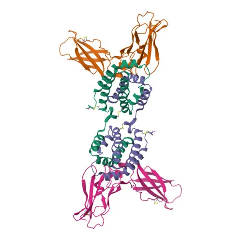
- Interleukin-10 receptor subunit alpha (IL-10RA), also designated CD210, IL-10R1, or CDw210a, is a transmembrane receptor belonging to the type II cytokine receptor family.
- It serves as the high-affinity ligand-binding subunit for the anti-inflammatory cytokine IL-10, playing a critical role in limiting excessive immune responses and tissue damage.
- Upon IL-10 binding, IL-10RA undergoes a conformational change that enables recruitment and binding of the IL-10RB co-receptor, forming a functional heterotetrameric signaling complex. Related references: PMID:16982608
- This complex activates the receptor-associated tyrosine kinases JAK1 and TYK2, which phosphorylate specific IL-10RA intracellular tyrosine residues, triggering the recruitment and phosphorylation of the transcription factor STAT3. Related references: PMID:12133952
- Phosphorylated STAT3 homodimerizes and translocates to the nucleus, where it induces anti-inflammatory gene expression. IL-10RA signaling also suppresses starvation-induced autophagy through STAT3. Related references: PMID:26962683
- IL-10RA is primarily expressed on the surface of hematopoietic cells, including B-cells, T-cells, NK cells, monocytes, and macrophages, but is absent from non-hematopoietic cells like fibroblasts and endothelial cells.
- The protein localizes to the cell membrane and cytoplasm, and it undergoes post-translational modifications such as glycosylation, phosphorylation, and ubiquitination.
Experimental Guidance and Technical Tips
- The immunogen corresponds to the extracellular domain (amino acids 22–235) of human IL-10RA, suggesting the antibody may recognize an epitope in this region and is suitable for surface staining of live cells.
- As an conjugate validated for flow cytometry, titration experiments should be considered to optimize the signal-to-noise ratio in the specific experimental system.
- Because IL-10RA expression is restricted to hematopoietic cells, include relevant human cell models (e.g., PBMCs, isolated immune cell subsets) and appropriate isotype controls in FC assays.
- Cell stimulation with IL-10 can influence receptor dynamics; for functional studies, consider comparing resting versus activated conditions.
CamelBio: Your One-Stop Sourcing Bridge
CamelBio is your integrated partner for IVD raw-material procurement, offering validated antibody pairs, optimized monoclonal antibodies, and bulk ancillary reagents—including rare-target sourcing if needed. For IL-10RA/CD210 research, we supply rabbit monoclonal antibodies (such as the conjugate highlighted here) to support immune-regulation studies, anti-inflammatory screening, and cell phenotyping. With comprehensive technical services and consulting available, CamelBio helps bridge your diagnostic development from concept to clinical application.
REQUEST A QUOTE
Our professional team will reply to you within one business day. Please feel free to contact us!