Monoclonal Antibodies
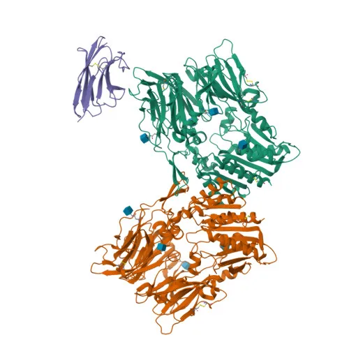
Rabbit anti-Human FAP Monoclonal Antibody for FC - Q12884
Item Number : CM0009346
Price varies based on specs and customizations
- Application
- FC
- Cross Reactivity
- Human
- Protein Weight
- 27kDa/87kDa

Shipping:
Contact us to get shipping details Enjoy On-time Dispatch Guarantee.
Why Choose Us
Easy ordering process, quality products, and dedicated support for your business success.
Core Product Specifications and Parameters
| Parameter | Value |
|---|---|
| Product Name | Rabbit anti-Human FAP mAb |
| Remarks/Alias | FAP; DPPIV; FAPA; FAPalpha; SIMP; prolyl endopeptidase FAP |
| Species | Human |
| GeneID (Human) | 2191 |
| GeneID | 2191 |
| Immunogen | Recombinant fusion protein containing a sequence corresponding to amino acids 26-760 of human FAP (NP_004451.2). |
| Source | Rabbit |
| Category | Monoclonal Antibodies |
| Application | FC |
| Cross Reactivity | Human |
| SWISS | Q12884 |
| Protein Weight | 27kDa/87kDa |
| Shipping | Ice bag |
Biological Background: Prolyl Endopeptidase FAP Function and Localization
- Prolyl endopeptidase FAP (FAP) is a cell surface glycoprotein serine protease, also known as fibroblast activation protein alpha, seprase, and 170 kDa melanoma membrane-bound gelatinase. Multiple isoforms exist due to alternative splicing.
- FAP exhibits both post-proline cleaving endopeptidase activity and dipeptidyl peptidase activity. The endopeptidase activity targets substrates with Ala/Ser-Gly-Pro-Ser/Asn/Ala sequences, such as alpha-2-antiplasmin and SPRY2. Related references: PMID:14751930, PMID:16223769, PMID:16410248
- The dipeptidyl peptidase activity hydrolyzes prolyl bonds in synthetic dipeptides and acts on natural neuropeptides including NPY, PYY, substance P, and BNP32. Related references: PMID:10347120, PMID:10593948, PMID:16175601, PMID:21314817
- FAP degrades gelatin and heat-denatured type I collagen, but not native collagen types I and IV, vitronectin, tenascin, laminin, fibronectin, fibrin, or casein, contributing to ECM remodeling. Related references: PMID:10347120, PMID:10455171, PMID:12376466
- Through its protease activities, FAP regulates pericellular proteolysis, promoting cell adhesion, migration, and invasion. It plays roles in tissue remodeling, fibrosis, wound healing, inflammation, and tumor progression, including enhancing angiogenesis and immune evasion.
- Subcellularly, FAP localizes to the cell surface and membrane, particularly in cell projections such as lamellipodia, invadopodia, and ruffles. Soluble forms are also secreted into the extracellular space and detected in the cytoplasm.
- Tissue expression is largely restricted to activated fibroblasts in pathological conditions: healing wounds, cirrhotic liver (hepatic stellate cells), inflamed synovial tissues, and reactive stroma of epithelial cancers. It is highly expressed in glioma cells and melanoma lines, but absent from normal liver. Adipose tissue also expresses FAP.
- Post-translational modifications include glycosylation and disulfide bond formation, and it is synthesized as a single-pass type II membrane protein with a signal-anchor.
Experimental Guidance and Technical Tips
- This antibody is designed for flow cytometry (FC) applications. The immunogen spans amino acids 26-760 of human FAP, covering the extracellular domain. The fluorophore allows direct detection without secondary reagents, reducing background.
- For cell surface staining of live cells, incubate with the antibody on ice in blocking buffer. For intracellular or total FAP detection, consider fixation and permeabilization protocols.
- Optimal antibody concentration should be determined by titration in the relevant cell system. Include appropriate isotype controls and viability dyes to exclude dead cells from analysis.
- Since FAP is expressed on activated fibroblasts in tumor stroma and healing tissues, use appropriate positive and negative controls (e.g., FAP-positive cancer-associated fibroblasts vs. normal tissue fibroblasts or FAP-negative cell lines).
- Due to potential shedding, secreted forms of FAP may be present in the supernatant; for accurate surface staining, wash cells thoroughly before staining.
CamelBio: Your One-Stop Sourcing Bridge
As a dedicated one-stop sourcing platform for IVD raw materials, CamelBio provides antibody pairs, monoclonal antibodies, and bulk ancillary reagents to streamline your assay development. This high-quality monoclonal antibody targeting FAP supports cancer and fibrosis diagnostic research. Our supply chain delivers validated antibodies with consistent lot-to-lot performance to meet your clinical and research needs.
REQUEST A QUOTE
Our professional team will reply to you within one business day. Please feel free to contact us!