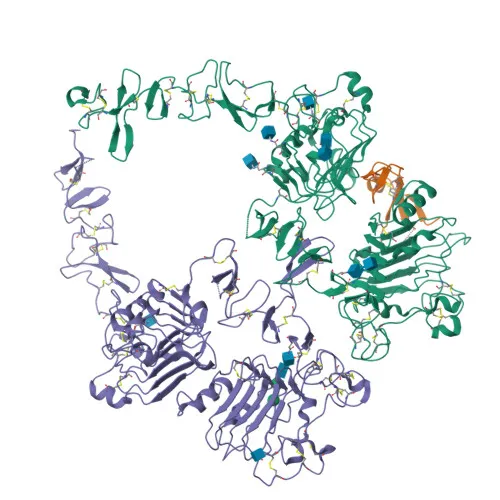
Monoclonal Antibodies
Rabbit anti-Human ErbB3/HER3 Monoclonal Antibody for Flow Cytometry - P21860
Item Number : CM0010819
Price varies based on specs and customizations
- Application
- Flow Cytometry (FC)
- Cross Reactivity
- Human
- Protein Weight
- 148 kDa

Shipping:
Contact us to get shipping details Enjoy On-time Dispatch Guarantee.
Why Choose Us
Easy ordering process, quality products, and dedicated support for your business success.
Core Product Specifications and Parameters
| Parameter | Value |
|---|---|
| Product Name | Rabbit anti-Human ErbB3/HER3 mAb |
| Remarks/Alias | HER3; FERLK; LCCS2; VSCN1; ErbB-3; c-erbB3; erbB3-S; MDA-BF-1; c-erbB-3; p180-ErbB3; p45-sErbB3; p85-sErbB3 |
| Species | Human |
| Gene ID | 2065 (human) |
| Immunogen | Recombinant fusion protein containing a sequence corresponding to amino acids 20-643 of human ErbB3/HER3 (NP_001973.2). |
| Source | Rabbit |
| Category | Monoclonal Antibodies |
| Application | Flow Cytometry (FC) |
| Cross Reactivity | Human |
| Swiss-Prot | P21860 |
| Protein Weight | 148 kDa |
| Shipping | Ice bag |
Biological Background: ErbB3/HER3 Function and Localization
- ErbB3 is a receptor tyrosine-protein kinase that functions as a cell surface receptor for neuregulins, primarily neuregulin-1 (NRG1). Ligand binding induces tyrosine phosphorylation and promotes association with the p85 subunit of phosphatidylinositol 3-kinase (PI3K). It may also be activated by CSPG5. Related references: PMID:20682778 PMID:15358134
- ErbB3 is involved in the regulation of myeloid cell differentiation, underscoring its role in hematopoietic lineage commitment. Related references: PMID:27416908
- The ERBB3 gene undergoes alternative splicing, generating multiple isoforms, including secreted variants that lack the transmembrane domain. This is consistent with its annotation as a secreted protein in addition to its membrane-bound form.
- Subcellularly, ErbB3 is predominantly localized to the cell membrane, with secreted isoforms found extracellularly. Tissue expression is notable in epithelial tissues and the brain.
- Post-translational modifications include glycosylation, disulfide bond formation, and phosphorylation on key tyrosine residues, which are critical for its signaling functions.
- As a member of the ERBB family of receptor tyrosine kinases, ErbB3 contains an ATP-binding domain and exhibits tyrosine kinase activity, though its kinase activity is reportedly weaker than other family members.
- Disease-associated variants of ERBB3 have been linked to conditions such as lethal congenital contracture syndrome 2 (LCCS2) and other disorders, highlighting its clinical relevance.
Experimental Guidance and Technical Tips
- The immunogen corresponds to the large extracellular domain (aa20-643) of human ErbB3, suggesting the antibody is likely to recognize the native cell surface receptor. For flow cytometry, consider using live, unfixed cells to maintain surface epitope integrity.
- Titrate the antibody to determine the optimal concentration for your specific application and include appropriate isotype controls and fluorescence-minus-one (FMO) controls.
- ErbB3 expression is reportedly prominent in epithelial tissues and certain brain regions. Validate in relevant cell lines (e.g., MCF-7, T47D) or primary samples known to express the target.
CamelBio: Your One-Stop Sourcing Bridge
CamelBio offers a streamlined, one-stop sourcing solution for IVD raw materials, supporting oncology and signal transduction research with products like this ErbB3/HER3 monoclonal antibody. From validated antibody pairs and optimized monoclonal antibodies to bulk ancillary reagents and rare-target sourcing, CamelBio accelerates your concept-to-clinic development with reliable procurement and technical support.
Product Datasheet
Rabbit anti-Human ErbB3/HER3 Monoclonal Antibody for Flow Cytometry - P21860
REQUEST A QUOTE
Our professional team will reply to you within one business day. Please feel free to contact us!