Monoclonal Antibodies
Rabbit anti-Human EGFR mAb for FC - P00533
Item Number : CM0012217
Price varies based on specs and customizations
- Application
- FC
- Cross Reactivity
- Human
- Protein Weight
- 44kDa/69kDa/77kDa/134kDa

Shipping:
Contact us to get shipping details Enjoy On-time Dispatch Guarantee.
Why Choose Us
Easy ordering process, quality products, and dedicated support for your business success.
Core Product Specifications and Parameters
| Parameter | Value |
|---|---|
| Product Name | Rabbit anti-Human EGFR mAb |
| Remarks/Alias | ERBB; ERRP; HER1; mENA; ERBB1; PIG61; NISBD2 |
| Species | Human |
| GeneID (Human) | 1956 |
| Immunogen | Recombinant protein | Recombinant fusion protein containing a sequence corresponding to amino acids 335-525 of human EGFR (NP_005219.2). |
| Source | Rabbit |
| Category | Monoclonal Antibodies |
| Application | FC |
| Cross Reactivity | Human |
| SWISS | P00533 |
| Protein Weight | 44kDa/69kDa/77kDa/134kDa |
| Shipping | Ice bag |
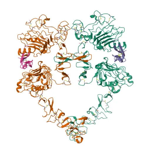
Biological Background: Epidermal Growth Factor Receptor Function and Localization
- EGFR is a receptor tyrosine kinase that binds EGF family ligands, triggering homo/heterodimerization, autophosphorylation, and activation of multiple downstream cascades including RAS-RAF-MEK-ERK, PI3K-AKT, PLCγ-PKC, and STAT modules; it also modulates NF-κB signaling and phosphorylates substrates like RGS16 and MUC1 to regulate cell migration and other processes. Related references: PMID:10805725 PMID:27153536 PMID:2790960 PMID:12297049 PMID:15611079 PMID:17909029 PMID:11116146 PMID:11602604 PMID:11483589 PMID:20462955
- EGFR functions as a receptor for hepatitis C virus (HCV), facilitating viral entry into hepatocytes by promoting CD81-CLDN1 receptor complex formation.
- Isoform 2 may act as an antagonist of EGF action, potentially modulating ligand-dependent signaling.
- Subcellularly, EGFR localizes to the cell membrane, endoplasmic reticulum membrane, Golgi membrane, nucleus membrane, endosome, and can be secreted; minor pools are found in the nucleus.
- EGFR is ubiquitously expressed across tissues, with isoform 2 also detected in ovarian cancers.
- Key post-translational modifications and features include phosphorylation, glycosylation, ubiquitination, and palmitoylation, as highlighted by UniProt keywords: host-virus interaction, proto-oncogene, tyrosine-protein kinase, membrane receptor, transmembrane, and signaling.
Experimental Guidance and Technical Tips
- The immunogen corresponds to the extracellular ligand-binding domain (aa 335–525) of human EGFR; the resulting rabbit monoclonal antibody likely recognizes native cell surface EGFR. For flow cytometry, perform antibody titration and include isotype controls to optimize staining conditions.
- This conjugate is suitable for live-cell surface staining; if intracellular detection is needed, validate appropriate fixation/permeabilization protocols for your cell type.
- Given EGFR’s broad tissue expression, confirm antibody specificity and signal-to-noise ratio in the desired biological sample (e.g., cancer cell lines, primary hepatocytes) under your experimental settings.
- For HCV entry studies, consider using the antibody to monitor EGFR expression on hepatocyte-derived lines (e.g., Huh7) and evaluating its role in CD81-CLDN1 complex formation.
CamelBio: Your One-Stop Sourcing Bridge
CamelBio provides a comprehensive portfolio of IVD-grade antibodies and assay-critical raw materials for EGFR-targeted diagnostic and research applications. From validated monoclonal antibodies for flow cytometry to optimized antibody pairs and bulk ancillary reagents, our one-stop sourcing platform supports every stage of diagnostic development. Partner with CamelBio for streamlined procurement, technical consultation, and scalable supply of EGFR-related raw materials tailored to your oncology or infectious disease IVD projects.
REQUEST A QUOTE
Our professional team will reply to you within one business day. Please feel free to contact us!