Monoclonal Antibodies
Rabbit anti-Human E-Cadherin/CD324 Monoclonal Antibody for Flow Cytometry (FC) - P12830
Item Number : CM0010384
Price varies based on specs and customizations
- Application
- FC
- Cross Reactivity
- Human
- Protein Weight
- 97kDa

Shipping:
Contact us to get shipping details Enjoy On-time Dispatch Guarantee.
Why Choose Us
Easy ordering process, quality products, and dedicated support for your business success.
Core Product Specifications and Parameters
| Parameter | Details |
|---|---|
| Product Name | Rabbit anti-Human E-Cadherin/CD324 mAb |
| Remarks/Alias | UVO; CDHE; ECAD; LCAM; Arc-1; BCDS1; CD324 |
| Species | Human |
| Gene ID (Human) | 999 |
| Gene ID | 999 |
| Immunogen | Recombinant protein (Recombinant fusion protein containing a sequence corresponding to amino acids 155-707 of human E-Cadherin/CD324 mAb(NP_004351.1)) |
| Source | Rabbit |
| Category | Monoclonal Antibodies |
| Application | FC (Flow Cytometry) |
| Cross Reactivity | Human |
| SWISS | P12830 |
| Protein Weight | 97kDa |
| Shipping | Ice bag |
Biological Background: E-Cadherin/Cadherin-1 Function and Localization
- Cadherin-1 (E-cadherin, CD324) is a calcium-dependent cell adhesion protein encoded by the CDH1 gene. It mediates homophilic cell–cell interactions, contributing to cell sorting and regulating epithelial cell mobility and proliferation. Related references: PMID:11976333
- Plays a role in desmosome formation by facilitating the recruitment of desmoglein-2 (DSG2) and desmoplakin (DSP) to desmosome plaques. Related references: PMID:29999492
- Promotes organization of radial actin fiber structure and cellular response to contractile forces via interaction with AMOTL2, anchoring actin fibers to CDH1 junction complexes at the cell membrane.
- Has a potent invasive suppressor role and serves as a ligand for integrin alpha-E/beta-7.
- The C-terminal fragment (E-Cad/CTF2) promotes non-amyloidogenic degradation of Abeta precursors, strongly inhibiting APP C99 and C83 production.
- Serves as a receptor for Listeria monocytogenes internalin A, mediating bacterial entry into host cells.
- Subcellular localization includes adherens junctions, desmosomes, cell membrane, endosomes, Golgi apparatus, and secreted forms. Tissue expression is prominent in epithelial cells of skin, esophagus, liver, and in granuloma macrophages.
- Undergoes alternative splicing and post-translational modifications including phosphorylation, glycosylation, and ubiquitination.
Experimental Guidance and Technical Tips
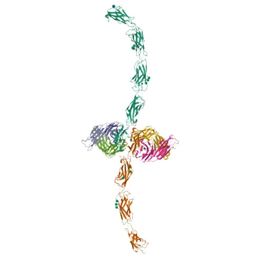
- The immunogen corresponds to amino acids 155–707 of human E-cadherin, spanning the majority of the extracellular domain. This design is expected to yield antibodies that recognize the surface-exposed protein, making the conjugate suitable for flow cytometry without cell permeabilization.
- For flow cytometry, consider using a cell line with confirmed E-cadherin expression (e.g., MCF-7, A431) and include an appropriate isotype control. Titrate the antibody to determine the optimal staining concentration for your specific experimental system.
- As the antibody is directly conjugated to , no secondary detection reagents are required, simplifying multiplex panels. Verify instrument compatibility with this fluorophore (excitation ~650 nm; emission ~668 nm).
CamelBio: Your One-Stop Sourcing Bridge
CamelBio provides diagnostic and life science manufacturers with streamlined access to critical raw materials. In addition to this E-cadherin/CD324 flow cytometry antibody, we supply validated antibody pairs, bulk-format monoclonal and polyclonal antibodies, ancillary reagents, and custom protein production services—all tailored to support your cell adhesion, oncology, and epithelial biology assay development. Our integrated sourcing model helps accelerate your projects from concept to clinical application.
Product Datasheet
Rabbit anti-Human E-Cadherin/CD324 Monoclonal Antibody for Flow Cytometry (FC) - P12830
REQUEST A QUOTE
Our professional team will reply to you within one business day. Please feel free to contact us!