Monoclonal Antibodies
Rabbit anti-Human Desmoglein 3/PVA Monoclonal Antibody for Flow Cytometry - P32926
Item Number : CM0010501
Price varies based on specs and customizations
- Application
- FC
- Cross Reactivity
- Human
- Protein Weight
- 108kDa

Shipping:
Contact us to get shipping details Enjoy On-time Dispatch Guarantee.
Why Choose Us
Easy ordering process, quality products, and dedicated support for your business success.
Core Product Specifications and Parameters
| Parameter | Value |
|---|---|
| Product Name | Rabbit anti-Human Desmoglein 3/PVA mAb |
| Remarks/Alias | PVA; ABOLM; CDHF6 |
| Species | Human |
| Gene ID (Human) | 1830 |
| Immunogen | Recombinant fusion protein containing a sequence corresponding to amino acids 50-615 of human Desmoglein 3/PVA (NP_001935.2). |
| Source | Rabbit |
| Category | Monoclonal Antibodies |
| Application | FC |
| Cross Reactivity | Human |
| SWISS | P32926 |
| Protein Weight | 108kDa |
| Shipping | Ice bag |
Biological Background: Desmoglein-3 Function and Localization
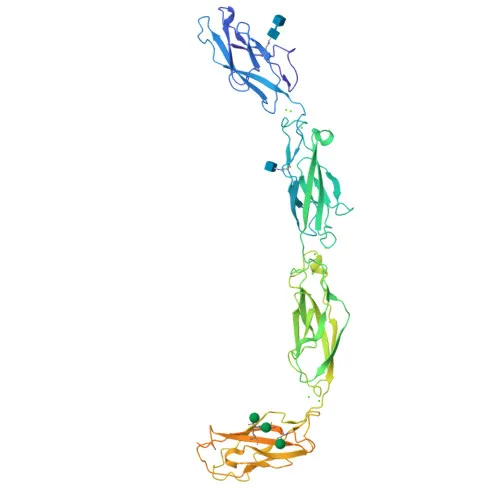
- Desmoglein-3 (DSG3), also termed 130 kDa pemphigus vulgaris antigen (PVA) or Cadherin family member 6 (CDHF6), is a calcium-binding transmembrane glycoprotein of the cadherin superfamily.
- DSG3 functions as a core desmosome component essential for strong intercellular adhesion, required for desmosome-mediated cell-cell attachment in keratinocytes and for anchoring telogen hair clubs in the follicle. Related references: PMID:31835537 PMID:9701552
- In keratinocytes, mechanical force triggers DSG3-dependent assembly of adherens and desmosome junctions; DSG3 also recruits YAP1 to the plasma membrane via a protein complex containing PKP1 and YWHAG, potentially modulating YAP1-mediated transcription and proliferation. Related references: PMID:31835537
- The cytoplasmic pool of DSG3 is critical for localizing CDH1 and CTNNB1 at developing adherens junctions and for maintaining stability of plaque components DSP, JUP, PKP1, PKP2, and PKP3. Related references: PMID:22294297
- DSG3 positively regulates cellular contractility and junction formation by organizing cortical F-actin bundles and anchoring actin to tight junctions via RAC1; it also restrains keratinocyte migration by suppressing p38MAPK signaling, potentially moderating wound healing. Related references: PMID:22796473 PMID:26763450
- The protein localizes to the cell membrane at desmosomes and tight junctions, with a cytoplasmic pool playing essential roles in junction remodeling.
- DSG3 is predominantly expressed in stratified epithelia—skin, oral and esophageal mucosa, and the outer root sheath of hair follicles—where it maintains epithelial barrier integrity.
Experimental Guidance and Technical Tips
- This -conjugated antibody is designed for flow cytometry and targets an extracellular epitope (amino acids 50–615). Consider using live, unpermeabilized cells for surface staining.
- Titrate the antibody to determine the optimal concentration for your sample type to achieve a good signal-to-noise ratio.
- Human keratinocyte lines (e.g., HaCaT) or epithelial cells with documented DSG3 expression are suitable positive controls. Validate expression in your target cells using available transcriptomic or proteomic data.
- Include appropriate isotype controls and fluorescence minus one (FMO) controls to ensure specific staining.
- Upon receipt, store the antibody at 2–8°C and protect from light. Avoid freeze-thaw cycles to maintain conjugate stability.
CamelBio: Your One-Stop Sourcing Bridge
CamelBio supports diagnostic developers, research institutes, and laboratories with a comprehensive portfolio of IVD raw materials, including monoclonal antibodies like this anti-Desmoglein 3 (DSG3) clone for flow cytometry. With reliable sourcing of validated antibody pairs, bulk reagents, and rare target raw materials, CamelBio serves as a single procurement gateway from concept to clinic.
Product Datasheet
Rabbit anti-Human Desmoglein 3/PVA Monoclonal Antibody for Flow Cytometry - P32926
REQUEST A QUOTE
Our professional team will reply to you within one business day. Please feel free to contact us!