Monoclonal Antibodies
Rabbit anti-Human CD97 Monoclonal Antibody for Flow Cytometry - P48960
Item Number : CM0011452
Price varies based on specs and customizations
- Application
- FC
- Cross Reactivity
- Human
- Protein Weight
- 81kDa/86kDa/92kDa

Shipping:
Contact us to get shipping details Enjoy On-time Dispatch Guarantee.
Why Choose Us
Easy ordering process, quality products, and dedicated support for your business success.
Core Product Specifications and Parameters
| Parameter | Value |
|---|---|
| Product Name | Rabbit anti-Human CD97 mAb |
| Remarks/Alias | CD97; TM7LN1 |
| Species | Human |
| Gene ID (Human) | 976 |
| Immunogen | Recombinant fusion protein containing a sequence corresponding to amino acids 21-398 of human CD97 (NP_510966.1) |
| Source | Rabbit |
| Category | Monoclonal Antibodies |
| Application | FC |
| Cross Reactivity | Human |
| SWISS | P48960 |
| Protein Weight | 81kDa/86kDa/92kDa |
| Shipping | Ice bag |
Biological Background: CD97 (ADGRE5) Function and Localization
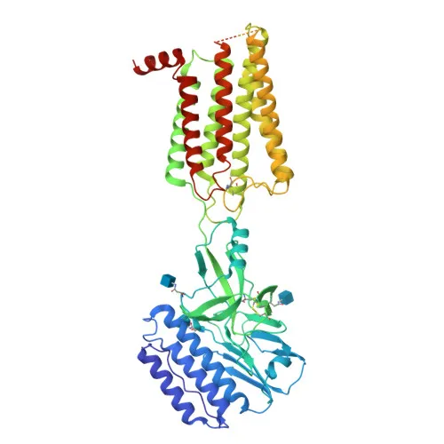
- CD97, also known as ADGRE5 or Leukocyte antigen CD97, is a member of the adhesion G protein-coupled receptor (GPCR) family and is involved in cell adhesion and signaling.
- It plays an essential role in leukocyte migration, mediating adhesion and signaling events crucial for immune cell trafficking.
- The receptor undergoes alternative splicing, producing isoforms with varying numbers of EGF-like domains, which may modulate ligand interactions.
- CD97 localizes to the cell membrane and can be shed into the extracellular space as a soluble form, consistent with its roles in adhesion and signaling.
- It is broadly expressed on hematopoietic cells, including activated lymphocytes, monocytes, macrophages, dendritic cells, and granulocytes, and is also abundant on smooth muscle cells.
- CD97 expression is upregulated in inflammatory conditions such as multiple sclerosis and rheumatoid arthritis, and soluble CD97 is detectable in synovial fluid.
- The protein contains multiple EGF-like domains, a GPCR proteolytic site, and a transmembrane helix, featuring disulfide bonds, glycosylation, and phosphorylation sites.
- Additionally, CD97 is expressed in various carcinomas (thyroid, colorectal, gastric, esophageal, pancreatic), suggesting a role in tumor biology.
Experimental Guidance and Technical Tips
- The immunogen corresponds to the extracellular region (amino acids 21–398) of human CD97, suggesting the antibody detects surface-expressed receptor, ideal for flow cytometry applications.
- For FC staining, consider titrating the antibody (e.g., 0.1–10 µg/mL) to optimize the signal-to-noise ratio in your specific cell type, and validate using appropriate positive (e.g., activated leukocytes) and negative controls.
- Since CD97 is shed into the extracellular space, be aware that soluble receptor in certain samples might compete for antibody binding; consider washing cells thoroughly before staining.
- Cross-reactivity is reported only for human; verify reactivity if using non-human primate cells due to potential sequence homology.
- The antibody is conjugated to , a far-red dye; ensure your flow cytometer has the appropriate laser and filter set (e.g., 633/635 nm excitation, 700–725 nm emission) for detection.
- Include appropriate isotype controls and viability dyes to gate on live, single cells for accurate analysis.
CamelBio: Your One-Stop Sourcing Bridge
As your comprehensive partner in IVD raw materials, CamelBio supports your CD97-related research and assay development with this high-quality -conjugated rabbit monoclonal antibody. We offer reliable supply of validated monoclonal and polyclonal antibodies, matched antibody pairs, and bulk ancillary reagents to streamline your workflow. For projects targeting rare or complex membrane proteins like adhesion GPCRs, CamelBio’s expert sourcing bridges the gap from concept to clinic.
Product Datasheet
Rabbit anti-Human CD97 Monoclonal Antibody for Flow Cytometry - P48960
REQUEST A QUOTE
Our professional team will reply to you within one business day. Please feel free to contact us!