Monoclonal Antibodies
Rabbit anti-Human CD97 mAb for FC - P48960
Item Number : CM0006065
Price varies based on specs and customizations
- Application
- FC
- Cross Reactivity
- Human
- Protein Weight
- 92kDa

Shipping:
Contact us to get shipping details Enjoy On-time Dispatch Guarantee.
Why Choose Us
Easy ordering process, quality products, and dedicated support for your business success.
Core Product Specifications and Parameters
| Parameter | Value |
|---|---|
| Product Name | Rabbit anti-Human CD97 mAb |
| Remarks/Alias | CD97; TM7LN1 |
| Species | Human |
| GeneID (Human) | 976 |
| GeneID | 976 |
| Immunogen | Recombinant protein|Recombinant fusion protein containing a sequence corresponding to amino acids 21-398 of human CD97 (NP_510966.1). |
| Source | Rabbit |
| Category | Monoclonal Antibodies |
| Application | FC |
| Cross Reactivity | Human |
| SWISS | P48960 |
| Protein Weight | 92kDa |
| Shipping | Ice bag |
Biological Background: CD97 Function and Localization
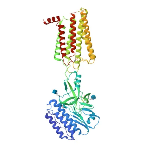
- CD97, also known as Adhesion G protein-coupled receptor E5 or Leukocyte antigen CD97, is a member of the adhesion GPCR family characterized by an extended extracellular N-terminus containing multiple EGF-like repeats.
- It functions as a receptor involved in both cell adhesion and signaling, playing an essential role in leukocyte migration.
- The protein is expressed as transmembrane and soluble isoforms; alternative splicing generates variants with different numbers of EGF-like domains.
- Subcellular localization: primarily found on the cell membrane, but also secreted into the extracellular space.
- Broadly expressed on most hematopoietic cells (activated lymphocytes, monocytes, macrophages, dendritic cells, granulocytes) and abundant in smooth muscle cells; also found in certain carcinomas (thyroid, colorectal, gastric, esophageal, pancreatic).
- Expression is upregulated under inflammatory conditions, such as in multiple sclerosis CNS and rheumatoid arthritis synovial tissue, with increased soluble CD97 detectable in synovial fluid.
- Post-translational modifications include glycosylation, disulfide bonds, and phosphorylation; the extracellular domain contains EGF-like calcium-binding sites.
Experimental Guidance and Technical Tips
- The immunogen used was a recombinant protein corresponding to amino acids 21-398 of human CD97, encompassing the extracellular domain. This design likely yields antibodies recognizing surface-accessible epitopes.
- For flow cytometry, use standard staining protocols with appropriate isotype controls; titrate the antibody to determine optimal working concentration.
- Validate reactivity in relevant human hematopoietic or carcinoma cell lines known to express CD97 (e.g., leukocytes, smooth muscle cell lines).
- Note that CD97 can be shed, so soluble receptor may compete in certain assays; consider using fresh cells or blocking with recombinant protein if necessary.
- Avoid relying on molecular weight determination by western blot as this antibody is recommended for FC only; verify specificity by staining cells with known CD97 expression patterns.
CamelBio: Your One-Stop Sourcing Bridge
CamelBio provides diagnostic manufacturers, laboratories, and research institutes with one-stop access to IVD raw materials, technical services, and consulting—covering every stage from concept to clinic. Our portfolio includes validated antibody pairs, optimized monoclonal/polyclonal antibodies, bulk ancillary reagents, and rare target raw-material sourcing for targets like CD97. Partner with CamelBio for seamless procurement of high-quality reagents to accelerate your immunology and oncology research projects.
REQUEST A QUOTE
Our professional team will reply to you within one business day. Please feel free to contact us!