Monoclonal Antibodies
Rabbit anti-Human CD9 Monoclonal Antibody () for Flow Cytometry - P21926
Item Number : CM0012670
Price varies based on specs and customizations
- Application
- FC
- Cross Reactivity
- Human
- Protein Weight
- 25kDa

Shipping:
Contact us to get shipping details Enjoy On-time Dispatch Guarantee.
Why Choose Us
Easy ordering process, quality products, and dedicated support for your business success.
Core Product Specifications and Parameters
| Parameter | Value |
|---|---|
| Product Name | Rabbit anti-Human CD9 mAb |
| Remarks/Alias | MIC3, MRP-1, BTCC-1, DRAP-27, TSPAN29, TSPAN-29 |
| Species | Human |
| Gene ID (Human) | 928 |
| Gene ID | 928 |
| Immunogen | Recombinant protein / Recombinant fusion protein containing amino acids 1-228 of human CD9 (NP_001760.1) |
| Source | Rabbit |
| Category | Monoclonal Antibodies |
| Application | FC |
| Cross Reactivity | Human |
| SWISS | P21926 |
| Protein Weight | 25kDa |
| Shipping | Ice bag |
Biological Background: CD9 Function and Localization
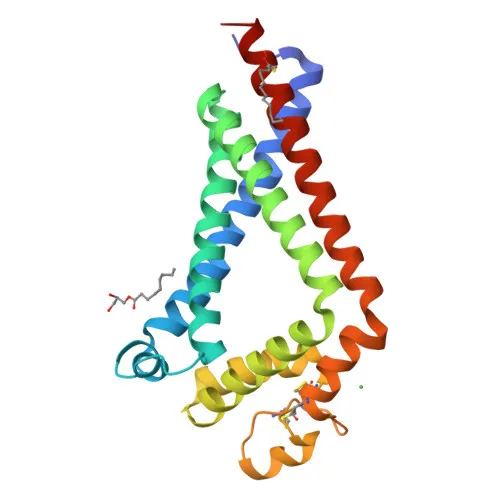
- CD9 is a member of the tetraspanin superfamily, also known as TSPAN29, p24, MRP-1, DRAP-27, BTCC-1, and leukocyte antigen MIC3. It functions as an integral membrane protein that associates with integrins and other tetraspanins.
- In oocytes, CD9 plays a critical role in sperm-egg fusion by organizing membrane microdomains and multiprotein complexes required for fertilization (By similarity).
- CD9 regulates platelet activation and aggregation through its association with integrins, contributing to hemostasis and thrombosis. Related references: PMID:18541721 PMID:8478605
- In myoblasts, CD9 associates with CD81 and PTGFRN to inhibit myotube fusion during muscle regeneration (By similarity).
- In macrophages, CD9 partners with CD81 and beta-1/beta-2 integrins to prevent cell fusion into multinucleated giant cells, modulating the immune response. Related references: PMID:12796480
- CD9 is involved in cell adhesion, motility, and tumor metastasis, influencing cancer progression and invasion. Related references: PMID:7511626 PMID:8478605
- Subcellularly, CD9 localizes to the cell membrane and is also found on secreted extracellular exosomes, facilitating intercellular communication.
- CD9 is abundantly expressed in platelets and a variety of hematopoietic and epithelial cells. Related references: PMID:19640571
Experimental Guidance and Technical Tips
- The immunogen covers the full extracellular domain of CD9 (amino acids 1-228), which may provide access to native epitopes on live cells, making it suitable for flow cytometry staining of surface CD9.
- For flow cytometry, consider titrating the antibody to determine the optimal concentration for your cell type, as CD9 expression levels can vary among hematopoietic and epithelial cells.
- Ensure proper isotype control and viability staining to distinguish CD9-positive populations accurately.
- Since CD9 is also found on extracellular exosomes, be aware that cell culture supernatants may contain shed CD9 that could interfere with staining; washing cells thoroughly before staining is recommended.
- The antibody is conjugated to , a bright and stable dye; avoid repeated freeze-thaw cycles and protect from light.
- Validate the antibody in the relevant sample system (e.g., human platelets, leukocytes, or cell lines) before use in quantitative assays.
CamelBio: Your One-Stop Sourcing Bridge
CamelBio connects diagnostic manufacturers and research laboratories with high-quality IVD raw materials, including the -conjugated anti-CD9 monoclonal antibody designed for flow cytometry applications targeting the tetraspanin CD9. Beyond this product, we offer one-stop procurement solutions covering validated antibody pairs, recombinant proteins, and ancillary reagents to accelerate your assay development from concept to clinic. Whether you need bulk quantities or rare target raw materials, CamelBio serves as your flexible sourcing bridge.
REQUEST A QUOTE
Our professional team will reply to you within one business day. Please feel free to contact us!