Monoclonal Antibodies
Rabbit anti-Human CD89 Monoclonal Antibody for FC - P24071
Item Number : CM0012544
Price varies based on specs and customizations
- Application
- FC
- Cross Reactivity
- Human
- Protein Weight
- 32kDa

Shipping:
Contact us to get shipping details Enjoy On-time Dispatch Guarantee.
Why Choose Us
Easy ordering process, quality products, and dedicated support for your business success.
Core Product Specifications and Parameters
| Parameter | Value |
|---|---|
| Product Name | Rabbit anti-Human CD89 mAb |
| Remarks/Alias | CD89; FcalphaR; FcalphaRI; CTB-61M7.2 |
| Species | Human |
| Gene ID (Human) | 2204 |
| Gene ID | 2204 |
| Immunogen | Recombinant protein|Recombinant fusion protein containing a sequence corresponding to amino acids 22-227 of human CD89 (NP_001991.1). |
| Source | Rabbit |
| Category | Monoclonal Antibodies |
| Application | FC |
| Cross Reactivity | Human |
| SWISS | P24071 |
| Protein Weight | 32kDa |
| Shipping | Ice bag |
Biological Background: CD89 Function and Localization
- CD89 (Immunoglobulin alpha Fc receptor), also known as FcalphaRI, is encoded by the FCAR gene in humans. It functions as a receptor for the Fc region of immunoglobulin alpha (IgA) and mediates immune effector functions such as cytokine production.
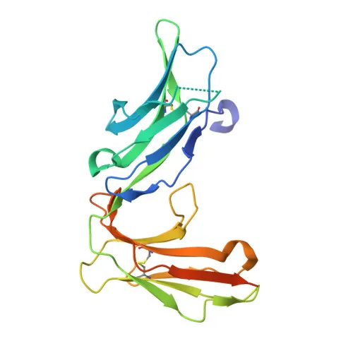
- The receptor is a member of the immunoglobulin superfamily, featuring two extracellular Ig-like domains, a transmembrane helix, and a short cytoplasmic tail. It contains disulfide bonds and is glycosylated, which is critical for IgA binding.
- Alternative splicing generates multiple isoforms (A.1, A.2, A.3) that exhibit differential expression patterns. These isoforms vary in their extracellular and cytoplasmic regions, potentially modulating signaling and trafficking.
- CD89 is primarily localized to the cell membrane of myeloid cells, including monocytes, macrophages, and neutrophils. Additionally, secreted forms have been detected, likely resulting from proteolytic cleavage or alternative splicing.
- Tissue-specific expression studies indicate that isoform A.1 and A.2 are present in alveolar macrophages, but only one isoform is expressed at the cell surface, suggesting intricate regulation of receptor presentation.
- As an IgA-binding protein, CD89 plays a role in mucosal immunity, linking humoral and cellular responses. Its engagement can trigger phagocytosis, respiratory burst, and release of inflammatory mediators.
Experimental Guidance and Technical Tips
- The immunogen comprises amino acids 22-227 of human CD89 (extracellular domain), so the resulting monoclonal antibody is expected to recognize native cell-surface CD89. Pre-adsorption or blocking with soluble CD89 may be considered to confirm specificity.
- For flow cytometry using this conjugate, gate on live single cells and use an appropriate isotype control antibody conjugated to the same fluorophore. Include Fc receptor blocking if working with myeloid cells to reduce non-specific binding.
- Given the expression of CD89 on monocytes and macrophages, human peripheral blood mononuclear cells (PBMCs) or myeloid cell lines like U937 or THP-1 (after treatment) are suitable positive control systems. Note that CD89 expression can vary with cell activation and differentiation stage.
- Secreted isoforms of CD89 may compete for antibody binding in certain sample types (e.g., serum or culture supernatants); where relevant, wash cells thoroughly before staining.
CamelBio: Your One-Stop Sourcing Bridge
CamelBio partners with diagnostic manufacturers, laboratories, and research institutes to provide comprehensive IVD raw-material solutions. For immune receptor targets like CD89, we offer validated monoclonal and polyclonal antibodies, optimized antibody pairs, and bulk ancillary reagents. From antigen sourcing to technical consulting, our one‑stop model supports every stage of assay development—helping you move from concept to clinic efficiently.
REQUEST A QUOTE
Our professional team will reply to you within one business day. Please feel free to contact us!