Monoclonal Antibodies
Rabbit anti-Human CD66a/CEACAM1 Monoclonal Antibody for FC - P13688
Item Number : CM0004499
Price varies based on specs and customizations
- Application
- FC
- Cross Reactivity
- Human
- Protein Weight
- 58kDa

Shipping:
Contact us to get shipping details Enjoy On-time Dispatch Guarantee.
Why Choose Us
Easy ordering process, quality products, and dedicated support for your business success.
Core Product Specifications and Parameters
| Parameter | Value |
|---|---|
| Product Name | Rabbit anti-Human CD66a/CEACAM1 mAb |
| Remarks/Alias | CEACAM1; BGP; BGP1; BGPI; carcinoembryonic antigen-related cell adhesion molecule 1 |
| Species | Human |
| GeneID (Human) | 634 |
| Immunogen | Recombinant fusion protein containing a sequence corresponding to amino acids 35-428 of human CD66a/CEACAM1 (NP_001703.2) |
| Source | Rabbit |
| Category | Monoclonal Antibodies |
| Application | FC |
| Cross Reactivity | Human |
| SWISS | P13688 |
| Protein Weight | 58kDa |
| Shipping | Ice bag |
Biological Background: CEACAM1 Function and Localization
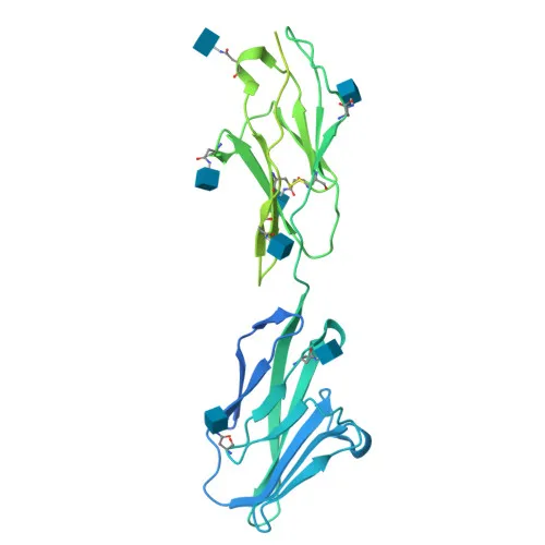
- CEACAM1 (Carcinoembryonic antigen-related cell adhesion molecule 1), also known as BGP-1, is a member of the carcinoembryonic antigen (CEA) immunoglobulin superfamily.
- It mediates calcium-independent homophilic cell adhesion, contributing to cell–cell interactions.
- CEACAM1 functions as a coinhibitory receptor in immune cells, suppressing TCR-mediated cytotoxicity and T cell proliferation by recruiting PTPN6 and dephosphorylating CD247 and ZAP70. Related references: PMID:18424730
- In natural killer (NK) cells, CEACAM1 inhibits KLRK1-mediated cytolysis through trans-homophilic binding and cis-interaction with KLRK1, leading to VAV1 dephosphorylation. Related references: PMID:23696226
- It negatively regulates neutrophil activity and IL1B production by facilitating a SYK-TLR4-CEACAM1 complex, and down-regulates neutrophil production via the CSF3R-STAT3 pathway.
- CEACAM1 also modulates insulin action by promoting INS clearance, regulates angiogenesis and vascular permeability, and inhibits cell migration through interaction with FLNA. Related references: PMID:16291724
- The protein is localized at the cell membrane (apical, basal, and lateral), cell junctions, adherens junctions, and can be secreted or found in cytoplasmic vesicles.
- It is expressed in colon epithelial cells, T cells, granulocytes, and lymphocytes, with leukocytes predominantly expressing isoforms containing a long cytoplasmic domain.
- Post-translational modifications include glycosylation, phosphorylation, and disulfide bond formation, contributing to its structural and signaling roles.
Experimental Guidance and Technical Tips
- The immunogen encompasses the N-terminal extracellular region (amino acids 35-428), ensuring reactivity against surface-exposed epitopes on live cells.
- This -conjugated antibody enables direct detection by flow cytometry without secondary reagents; consider using appropriate isotype controls and Fc receptor blocking steps.
- For optimal staining, titrate the antibody in your specific cell system and validate performance under your experimental conditions.
- Given its role in immune regulation, this antibody may be useful for studying CEACAM1 in T cells, NK cells, and tumor cell lines.
CamelBio: Your One-Stop Sourcing Bridge
CamelBio offers a comprehensive portfolio of IVD raw materials, including validated antibodies and antigens for immune-oncology and cell adhesion research. For your CEACAM1/CD66a detection needs, we provide conjugated monoclonal antibodies, bulk reagents, and custom sourcing solutions. Partner with us to accelerate your assay development from target selection to clinical application.
REQUEST A QUOTE
Our professional team will reply to you within one business day. Please feel free to contact us!