Monoclonal Antibodies
Rabbit anti-Human CD6 mAb for FC - P30203
Item Number : CM0010733
Price varies based on specs and customizations
- Application
- FC
- Cross Reactivity
- Human
- Protein Weight
- 58kDa/60kDa/63kDa/64kDa/68kDa/71kDa

Shipping:
Contact us to get shipping details Enjoy On-time Dispatch Guarantee.
Why Choose Us
Easy ordering process, quality products, and dedicated support for your business success.
Core Product Specifications and Parameters
| Parameter | Value |
|---|---|
| Product Name | Rabbit anti-Human CD6 mAb |
| Remarks/Alias | CD6; TP120; CD6 molecule |
| Species | Human |
| Gene ID (Human) | 923 |
| Gene ID | 923 |
| Immunogen | Recombinant fusion protein containing a sequence corresponding to amino acids 18-364 of human CD6 (NP_006716.3) |
| Source | Rabbit |
| Category | Monoclonal Antibodies |
| Application | FC |
| Cross Reactivity | Human |
| SWISS | P30203 |
| Protein Weight | 58kDa/60kDa/63kDa/64kDa/68kDa/71kDa |
| Shipping | Ice bag |
Biological Background: CD6 Function and Localization
- CD6 (T-cell differentiation antigen CD6, also known as T12/TP120) is a cell adhesion molecule that mediates cell-cell contacts through interaction with ALCAM/CD166, regulating T-cell responses. It contributes to signaling cascades triggered by the TCR/CD3 complex and promotes the formation and maturation of the immunological synapse. Related references: PMID:15048703 PMID:15294938 PMID:16352806 PMID:24584089
- CD6 functions as a costimulatory molecule, enhancing T-cell activation and proliferation, and is involved in cytokine production. Related references: PMID:15294938 PMID:16352806 PMID:16914752
- CD6 serves as a calcium-dependent pattern recognition receptor binding both Gram-positive and Gram-negative bacteria. It recognizes lipopolysaccharide (LPS) and lipoteichoic acid, triggering activation of MAP kinase signaling cascades and pro-inflammatory cytokine secretion. Related references: PMID:17601777
- CD6 is widely expressed on immune cells, including thymocytes, peripheral blood T-cells, natural killer (NK) cells, and a subset of B-1 cells. Soluble CD6 is detectable in blood serum.
- CD6 is a single-pass type I membrane protein, with subcellular localization at the cell membrane; a secreted form is also produced.
- Alternative splicing generates multiple CD6 isoforms with predicted molecular weights ranging from 58 to 71 kDa, reflected in multiple bands by western blot.
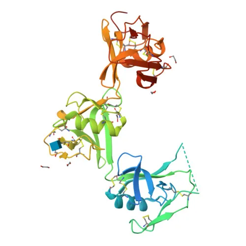
- The CD6 protein contains multiple scavenger receptor cysteine-rich (SRCR) domains and undergoes post-translational modifications including glycosylation and phosphorylation, consistent with its functions in innate and adaptive immunity.
Experimental Guidance and Technical Tips
- For flow cytometry, titrate the antibody to determine the optimal working concentration for your specific cell type and experimental conditions.
- Use human PBMCs or T-cell lines (e.g., Jurkat) as positive controls, and ensure Fc receptor blocking to minimize non-specific binding.
- Co-staining with anti-CD3 may help identify T-cell subsets; consider using isotype controls for accurate gating.
- Since CD6 is expressed on NK cells and some B-1 cells, broaden your panel to exclude these populations if necessary.
- Validate the antibody in your target sample system, as glycosylation and isoform diversity could affect epitope recognition.
CamelBio: Your One-Stop Sourcing Bridge
CamelBio offers a comprehensive portfolio of IVD raw materials, including validated antibody pairs, optimized monoclonal and polyclonal antibodies, and bulk ancillary reagents. This anti-CD6 monoclonal antibody supports immunology and inflammation research, complementing our one-stop procurement services from concept to clinic. Leverage our expertise to streamline your diagnostic development with reliable, high-performance raw materials.
REQUEST A QUOTE
Our professional team will reply to you within one business day. Please feel free to contact us!