Monoclonal Antibodies
Rabbit anti-Human CD59 Monoclonal Antibody for Flow Cytometry - P13987
Item Number : CM0008666
Price varies based on specs and customizations
- Application
- FC
- Cross Reactivity
- Human
- Protein Weight
- 14kDa

Shipping:
Contact us to get shipping details Enjoy On-time Dispatch Guarantee.
Why Choose Us
Easy ordering process, quality products, and dedicated support for your business success.
Core Product Specifications and Parameters
| Parameter | Value |
|---|---|
| Product Name | Rabbit anti-Human CD59 mAb |
| Remarks/Alias | 1F5; EJ16; EJ30; EL32; G344; MIN1; MIN2; MIN3; MIRL; HRF20; MACIF; MEM43; MIC11; MSK21; 16.3A5; HRF-20; MAC-IP; p18-20 |
| Species | Human |
| GeneID (Human) | 966 |
| GeneID | 966 |
| Immunogen | Recombinant protein |
| Source | Rabbit |
| Category | Monoclonal Antibodies |
| Application | FC |
| Cross Reactivity | Human |
| SWISS | P13987 |
| Protein Weight | 14kDa |
| Shipping | Ice bag |
Biological Background: CD59 Function and Localization
- CD59 glycoprotein, also known as protectin, MAC-inhibitory protein (MAC-IP), membrane inhibitor of reactive lysis (MIRL), or HRF-20, is a key regulator of the complement system.
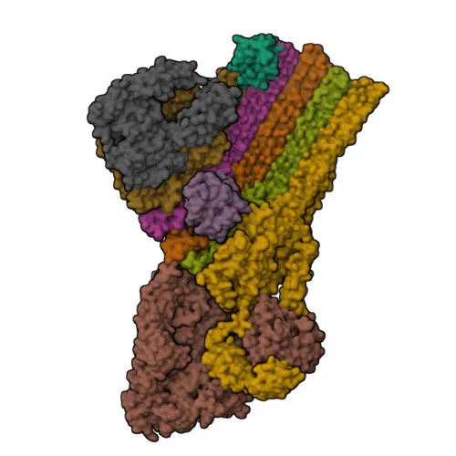
- CD59 is a potent inhibitor of the complement membrane attack complex (MAC) and protects human cells from complement-mediated lysis. It binds to the beta-hairpins of C8 (C8A and C8B) in the assembling MAC, forming an intermolecular beta-sheet that prevents incorporation of multiple copies of C9 required for complete formation of the osmolytic pore. Related references: PMID:11882685, PMID:1698710, PMID:2475111, PMID:36797260
- The soluble form of CD59, found in urine, retains specific complement binding activity but exhibits greatly reduced ability to inhibit MAC assembly on cell membranes.
- CD59 is primarily localized to the cell membrane via a glycosylphosphatidylinositol (GPI) anchor but can also be secreted.
- Post-translational modifications include N-glycosylation, disulfide bond formation, and glycation, contributing to its structural stability and function.
- Mutations in CD59 are associated with hereditary hemolytic anemia.
- Alternative splicing of the CD59 gene generates multiple isoforms.
Experimental Guidance and Technical Tips
- The immunogen corresponds to the N-terminal extracellular region of CD59 (aa 26–101), which is exposed on the cell surface; the conjugate is suitable for direct flow cytometry detection of native CD59 without a secondary antibody.
- For flow cytometry, titrate the antibody to determine the optimal staining concentration for your specific cell type; excess antibody may increase background.
- Include appropriate isotype controls and viability dyes to distinguish specific membrane staining from non-specific binding to dead cells.
- CD59 expression can vary among cell types and activation states; validate the staining in the relevant biological system (e.g., peripheral blood mononuclear cells, tumor cell lines) and consider co-staining with other complement regulatory markers.
CamelBio: Your One-Stop Sourcing Bridge
CamelBio streamlines the procurement of IVD raw materials, offering this anti-CD59 antibody as part of a comprehensive portfolio for complement research and immuno-oncology applications. For diagnostic manufacturers, CamelBio provides validated antibody pairs, optimized monoclonal/polyclonal antibodies, bulk ancillary reagents, and specialized rare target raw-material sourcing to support assay development. Our integrated supply chain and technical support help accelerate your projects from concept to clinic.
Product Datasheet
Rabbit anti-Human CD59 Monoclonal Antibody for Flow Cytometry - P13987
REQUEST A QUOTE
Our professional team will reply to you within one business day. Please feel free to contact us!