Monoclonal Antibodies
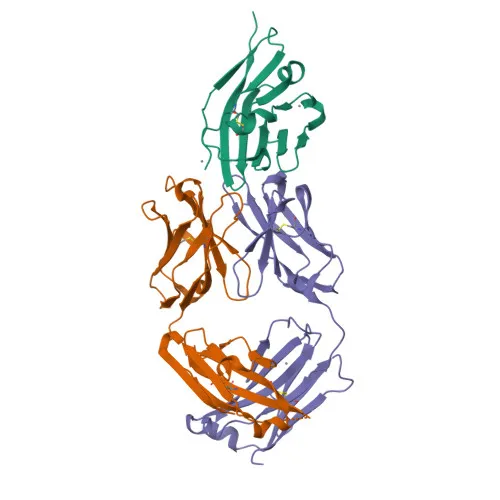
Rabbit anti-Human CD47 Monoclonal Antibody for FC - Q08722
Item Number : CM0004261
Price varies based on specs and customizations
- Application
- FC
- Cross Reactivity
- Human
- Protein Weight
- 31kDa/33kDa/35kDa

Shipping:
Contact us to get shipping details Enjoy On-time Dispatch Guarantee.
Why Choose Us
Easy ordering process, quality products, and dedicated support for your business success.
Core Product Specifications and Parameters
| Parameter | Value |
|---|---|
| Product Name | Rabbit anti-Human CD47 mAb |
| Remarks/Alias | CD47; IAP; MER6; OA3; CD47 molecule |
| Species | Human |
| Gene ID | 961 |
| Immunogen | Recombinant fusion protein containing a sequence corresponding to amino acids 19-139 of human CD47 (NP_001768.1) |
| Source | Rabbit |
| Category | Monoclonal Antibodies |
| Application | FC |
| Cross Reactivity | Human |
| SWISS | Q08722 |
| Protein Weight | 31kDa/33kDa/35kDa |
| Shipping | Ice bag |
Biological Background: CD47 Function and Localization
-
CD47 acts as an adhesive protein mediating cell-to-cell interactions and as a receptor for thrombospondin-1 (THBS1), thereby modulating integrin signaling and downstream heterotrimeric G protein activation. Related references: PMID:11509594 PMID:15383453 PMID:19004835 PMID:7691831 PMID:8550562
-
CD47 is involved in multiple physiological and pathological processes including signal transduction, cardiovascular homeostasis, inflammation, apoptosis, angiogenesis, cellular self-renewal, and immunoregulation. Related references: PMID:11509594 PMID:15383453 PMID:19004835
-
CD47 functions as a receptor for signal regulatory protein alpha (SIRPA), inhibiting dendritic cell maturation and cytokine production, and for SIRPG, mediating T-cell co-stimulation and proliferation. Related references: PMID:11509594 PMID:15383453
-
CD47 modulates pulmonary endothelin EDN1 signaling and contributes to hippocampal memory and synaptic plasticity. Related references: PMID:27742621
-
CD47 suppresses angiogenesis and may be involved in aging-related metabolic dysregulation, while also negatively modulating wound healing and inhibiting stem cell self-renewal in response to THBS1. Related references: PMID:32679764
-
CD47 is primarily a cell membrane protein, consistent with its role in adhesion and signaling.
-
CD47 is broadly expressed in normal adult tissues and overexpressed in ovarian tumors, with high abundance in epithelial cells, brain, and macrophages. Related references: PMID:39121194
-
Key post-translational modifications and structural features include glycosylation, disulfide bonds, an immunoglobulin-like domain, and alternative splicing, reflecting its dynamic regulatory roles.
Experimental Guidance and Technical Tips
- The immunogen corresponds to amino acids 19-139 of human CD47, which covers the extracellular IgV-like domain. This design likely targets the functionally relevant ligand-binding region; consider this when designing blocking or neutralization assays.
- This antibody is conjugated to , a bright and photostable fluorophore suitable for flow cytometry. Optimize staining concentration and ensure proper compensation in multicolor panels.
- Validate antibody performance in the relevant sample type (e.g., human PBMCs, cell lines) and include appropriate isotype controls and FcR blocking to minimize non-specific binding.
- For alternative applications such as immunofluorescence or immunohistochemistry, additional validation may be required due to the conjugate and clone specificity.
CamelBio: Your One-Stop Sourcing Bridge
As a trusted IVD raw material partner, CamelBio supports diagnostic developers with high-quality reagents such as this anti-CD47 monoclonal antibody for immune-oncology and hematology flow cytometry applications. CamelBio offers one-stop access to validated antibodies, bulk ancillary reagents, and custom sourcing for rare targets to accelerate your assay development from research to clinic.
REQUEST A QUOTE
Our professional team will reply to you within one business day. Please feel free to contact us!