Monoclonal Antibodies
Rabbit anti-Human CD33 Monoclonal Antibody for Flow Cytometry - P20138
Item Number : CM0004807
Price varies based on specs and customizations
- Application
- FC
- Cross Reactivity
- Human
- Protein Weight
- 25kDa/33kDa/39kDa

Shipping:
Contact us to get shipping details Enjoy On-time Dispatch Guarantee.
Why Choose Us
Easy ordering process, quality products, and dedicated support for your business success.
Core Product Specifications and Parameters
| Parameter | Value |
|---|---|
| Product Name | Rabbit anti-Human CD33 mAb |
| Remarks/Alias | CD33; SIGLEC-3; SIGLEC3; p67; CD33 molecule |
| Species | Human |
| Gene ID (Human) | 945 |
| Gene ID | 945 |
| Immunogen | Recombinant fusion protein containing a sequence corresponding to amino acids 18-259 of human CD33(NP_001763.3) |
| Source | Rabbit |
| Category | Monoclonal Antibodies |
| Application | FC |
| Cross Reactivity | Human |
| SWISS | P20138 |
| Protein Weight | 25kDa/33kDa/39kDa |
| Shipping | Ice bag |
Biological Background: CD33 Function and Localization
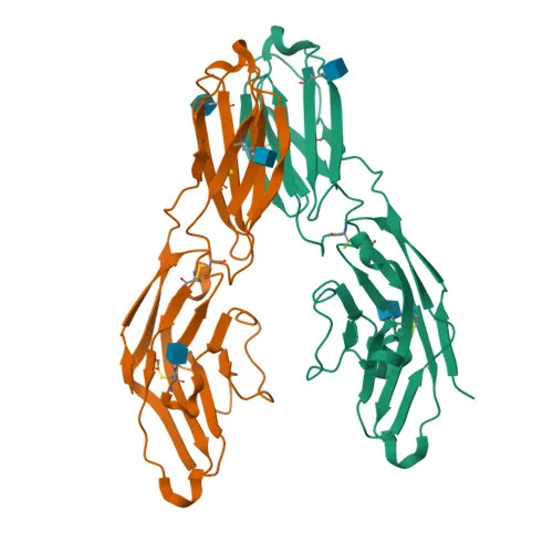
- Myeloid cell surface antigen CD33 (Siglec-3, gp67) is a type 1 transmembrane receptor belonging to the sialic acid-binding immunoglobulin-like lectin (Siglec) family. It is encoded by the CD33 gene.
- CD33 mediates cell-cell interactions and maintains immune cells in a resting state by preferentially recognizing and binding alpha-2,3- and alpha-2,6-linked sialic acid-bearing glycans. Related references: PMID:10611343, PMID:11320212, PMID:15597323, PMID:7718872
- Upon ligand engagement (e.g., C1q or sialylated glycoproteins), immunoreceptor tyrosine-based inhibitory motifs (ITIMs) in the cytoplasmic tail are phosphorylated by Src-like kinases such as LCK, creating docking sites for the protein-tyrosine phosphatases PTPN6/SHP-1 and PTPN11/SHP-2. Related references: PMID:10887109, PMID:28325905, PMID:10206955, PMID:10556798
- The recruited phosphatases dephosphorylate downstream signaling molecules, with one repressive effect depending on phosphoinositide 3-kinase/PI3K. Related references: PMID:15597323
- CD33 is predominantly expressed on the surface of monocytic and myeloid lineage cells (monocytes, macrophages, granulocytes) and, in the brain, on microglial cells.
- Subcellularly, CD33 localizes to the cell membrane and peroxisomes. It contains a signal peptide, an immunoglobulin-like V-type domain, and a transmembrane helix.
- The protein undergoes alternative splicing and post-translational modifications including phosphorylation, N-linked glycosylation, and disulfide bond formation. The predicted molecular weight is approximately 39.8 kDa, but multiple bands (25 kDa, 33 kDa, 39 kDa) are observed due to differential glycosylation and isoform expression.
Experimental Guidance and Technical Tips
- The immunogen (amino acids 18-259) encompasses the entire extracellular domain of human CD33, favoring recognition of the native, cell-surface form of the receptor.
- For flow cytometry, titrate the antibody to determine the optimal concentration. Use known CD33-positive cell lines (e.g., THP-1, U937) or human peripheral blood monocytes as positive controls.
- The conjugate allows direct detection without secondary reagents. Protect the antibody from light and avoid repeated freeze-thaw cycles to maintain fluorescence intensity.
- If intracellular staining is required (e.g., for peroxisomal localization), consider fixation and permeabilization protocols compatible with the fluorophore.
CamelBio: Your One-Stop Sourcing Bridge
CamelBio supports diagnostic manufacturers and research institutions with reliable access to high-quality IVD raw materials, including validated monoclonal antibodies such as this CD33 clone. Our portfolio covers antibody pairs, optimized monoclonal/polyclonal antibodies, bulk ancillary reagents, and rare target sourcing—streamlining your development from concept to clinic. Let CamelBio be your partner in efficient procurement and technical consultation.
Product Datasheet
Rabbit anti-Human CD33 Monoclonal Antibody for Flow Cytometry - P20138
REQUEST A QUOTE
Our professional team will reply to you within one business day. Please feel free to contact us!