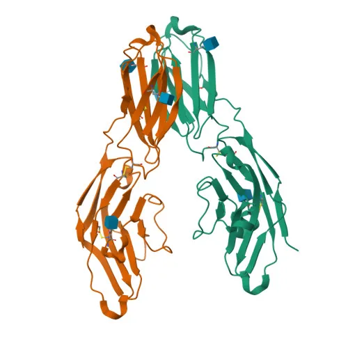
Monoclonal Antibodies
Rabbit anti-Human CD33 mAb for FC - P20138
Item Number : CM0009162
Price varies based on specs and customizations
- Application
- FC
- Cross Reactivity
- Human
- Protein Weight
- 40kDa

Shipping:
Contact us to get shipping details Enjoy On-time Dispatch Guarantee.
Why Choose Us
Easy ordering process, quality products, and dedicated support for your business success.
Core Product Specifications and Parameters
| Parameter | Value |
|---|---|
| Product Name | Rabbit anti-Human CD33 mAb |
| Remarks/Alias | p67; SIGLEC3; SIGLEC-3 |
| Species | Human |
| GeneID (human) | 945 |
| GeneID | 945 |
| Immunogen | Recombinant protein |
| Source | Rabbit |
| Category | Monoclonal Antibodies |
| Application | FC |
| Cross Reactivity | Human |
| SWISS | P20138 |
| Protein Weight | 40kDa |
| Shipping | Ice bag |
Biological Background: CD33 Function and Localization
- CD33, also known as Siglec-3 (sialic acid-binding Ig-like lectin 3) or p67, is a transmembrane receptor belonging to the immunoglobulin superfamily and Siglec family of sialic acid-binding lectins. It is primarily expressed on cells of the myeloid lineage.
- CD33 preferentially binds alpha-2,3- and alpha-2,6-linked sialic acid-bearing glycans, mediating cell-cell interactions and maintaining immune cells in a resting state. Ligand engagement triggers phosphorylation of its cytoplasmic immunoreceptor tyrosine-based inhibitory motifs (ITIMs) by Src-like kinases such as LCK, creating docking sites for the protein-tyrosine phosphatases SHP-1 and SHP-2. These phosphatases then dephosphorylate downstream signaling molecules to suppress cellular activation. Related references: PMID:7718872, PMID:10887109, PMID:28325905, PMID:10206955, PMID:10556798, PMID:10611343, PMID:11320212
- The inhibitory function of CD33 on monocyte activation requires phosphoinositide 3-kinase (PI3K) activity. Related references: PMID:15597323
- CD33 is localized to the cell membrane and peroxisomes, as indicated by subcellular fractionation and immunofluorescence studies.
- The protein is predominantly expressed on monocytic/myeloid lineage cells; in the brain, it is mainly expressed on microglial cells, where it has been implicated in neurodegenerative diseases.
- Post-translational modifications include N-linked glycosylation, disulfide bond formation, and phosphorylation of ITIM tyrosine residues. Alternative splicing generates multiple isoforms.
Experimental Guidance and Technical Tips
- The immunogen used for this antibody corresponds to the extracellular domain (amino acids 18-259) of human CD33. This region is exposed on the cell surface, making it suitable for live-cell flow cytometry without permeabilization.
- is a green-fluorescent dye with excitation/emission maxima of approximately 491/516 nm, compatible with the FITC channel on most flow cytometers. Ensure proper compensation if using in multicolor panels.
- For optimal staining, titrate the antibody to determine the working concentration for your specific experimental system. Include appropriate isotype control and fluorescence minus one (FMO) controls to set gating thresholds.
- Because CD33 expression levels can vary with cell type and activation state, validate the antibody in your sample matrix (e.g., peripheral blood mononuclear cells, bone marrow, or cell lines).
CamelBio: Your One-Stop Sourcing Bridge
CamelBio is a dedicated provider of IVD raw materials, supporting diagnostic manufacturers and researchers with validated antibodies, bulk reagents, and tailored sourcing solutions. For myeloid cell marker analysis in hematology and immunology workflows, our portfolio includes CD33 antibodies in various formats, alongside antibody pairs, recombinant proteins, and ancillary reagents to streamline your assay development. Contact us to learn how we can assist from concept to clinical application.
REQUEST A QUOTE
Our professional team will reply to you within one business day. Please feel free to contact us!