Monoclonal Antibodies
Rabbit anti-Human CD273/PD-L2 mAb for Flow Cytometry - Q9BQ51
Item Number : CM0011814
Price varies based on specs and customizations
- Application
- FC
- Cross Reactivity
- Human
- Protein Weight
- 21kDa/31kDa

Shipping:
Contact us to get shipping details Enjoy On-time Dispatch Guarantee.
Why Choose Us
Easy ordering process, quality products, and dedicated support for your business success.
Core Product Specifications and Parameters
| Parameter | Description |
|---|---|
| Product Name | Rabbit anti-Human CD273/PD-L2 mAb |
| Remarks/Alias | B7DC; Btdc; PDL2; CD273; PD-L2; PDCD1L2; bA574F11.2; PDCD1LG2 |
| Species | Human |
| Gene ID (Human) | 80380 |
| Gene ID | 80380 |
| Immunogen | Recombinant fusion protein containing a sequence corresponding to amino acids 20-219 of human CD273/PD-L2 (NP_079515.2) |
| Source | Rabbit |
| Category | Monoclonal Antibodies |
| Application | FC |
| Cross Reactivity | Human |
| SWISS | Q9BQ51 |
| Protein Weight | 21kDa/31kDa |
| Shipping | Ice bag |
Biological Background: PD-L2 (CD273) Function and Localization
- PD-L2 (CD273, B7-DC) is a type I transmembrane glycoprotein and member of the B7 family of immune regulatory ligands.
- It negatively regulates T-cell responses by binding to the PD-1 receptor, leading to suppression of T-cell proliferation, cell cycle arrest, and reduced cytokine production (PubMed:11224527). Related references: PMID:11224527
- PD-L2 is critically involved in the induction and maintenance of immune tolerance to self, thereby preventing autoimmunity (PubMed:11224527, PubMed:12538684). Related references: PMID:11224527, PMID:12538684
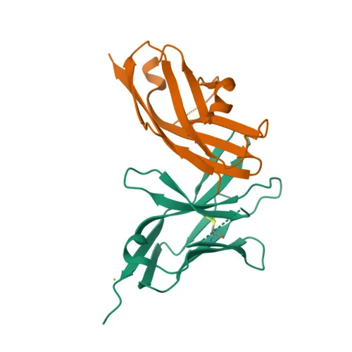
- Its protein structure includes an extracellular IgV-like domain, a transmembrane helix, and a short cytoplasmic tail; alternative splicing produces both membrane-anchored and soluble secreted isoforms.
- Subcellularly, PD-L2 is found on the cell surface, within the endomembrane system, and as a secreted protein, consistent with its role in both juxtacrine and paracrine signaling.
- It is highly expressed in non-lymphoid tissues like heart, placenta, pancreas, lung, and liver, with lower expression in lymphoid organs (spleen, lymph nodes, thymus), suggesting a role in peripheral tolerance.
- PD-L2 signaling is essential for maintaining immune homeostasis and preventing autoimmune responses.
Experimental Guidance and Technical Tips
- The immunogen corresponds to the extracellular domain (a.a. 20-219), encompassing the IgV-like domain critical for PD-1 binding. This design may favor detection of the native receptor-binding conformation in flow cytometry.
- For FC applications, consider titrating the antibody and using appropriate isotype controls to assess background staining, as PD-L2 expression levels can vary among different cell types.
- Due to the existence of a secreted form, cell surface staining might be affected by competitive shedding or internalization; validate in the relevant sample system.
- PD-L2 expression can be induced by inflammatory cytokines; thus, stimulation conditions may influence detection.
- This antibody is specific for human PD-L2; cross-species reactivity should be verified if used in non-human models.
CamelBio: Your One-Stop Sourcing Bridge
CamelBio provides diagnostic manufacturers and research labs with integrated access to IVD antibodies, reagents, and technical support—from early-stage development to clinical applications. This anti-PD-L2 monoclonal antibody supports immune checkpoint research and flow cytometry-based diagnostics development. Leverage CamelBio’s portfolio of validated antibody pairs, bulk reagents, and sourcing capabilities for rare targets to accelerate your biomarker discovery and assay development.
REQUEST A QUOTE
Our professional team will reply to you within one business day. Please feel free to contact us!