Monoclonal Antibodies
Rabbit anti-Human CD261/TRAIL-R1 Monoclonal Antibody for Flow Cytometry - O00220
Item Number : CM0010827
Price varies based on specs and customizations
- Application
- FC
- Cross Reactivity
- Human
- Protein Weight
- 50kDa

Shipping:
Contact us to get shipping details Enjoy On-time Dispatch Guarantee.
Why Choose Us
Easy ordering process, quality products, and dedicated support for your business success.
Core Product Specifications and Parameters
| Parameter | Value |
|---|---|
| Product Name | Rabbit anti-Human CD261/TRAIL-R1 mAb |
| Remarks/Alias | TNFRSF10A; APO2; CD261; DR4; TRAILR-1; TRAILR1; TNF receptor superfamily member 10a |
| Species | Human |
| Gene ID (Human) | 8797 |
| Gene ID | 8797 |
| Immunogen | Recombinant fusion protein containing a sequence corresponding to amino acids 124-232 of human CD261/TRAIL-R1 (NP_003835.3) |
| Source | Rabbit |
| Category | Monoclonal Antibodies |
| Application | FC |
| Cross Reactivity | Human |
| SWISS | O00220 |
| Protein Weight | 50kDa |
| Shipping | Ice bag |
Biological Background: CD261/TRAIL-R1 Function and Localization
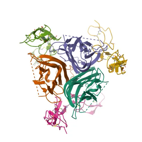
- TNFRSF10A, also known as Death receptor 4 (DR4) or TRAIL-R1, is a member of the tumor necrosis factor receptor superfamily. It serves as a receptor for the cytotoxic ligand TNFSF10/TRAIL. Related references: PMID:26457518, PMID:38532423
- Upon TRAIL binding, the receptor recruits the adapter molecule FADD and caspase-8 to form the death-inducing signaling complex (DISC), which proteolytically activates caspase-8 and initiates the apoptotic caspase cascade. Related references: PMID:19090789
- TRAIL-R1 also promotes the activation of NF-kappa-B, linking it to inflammatory and survival signaling pathways. Related references: PMID:9430227
- The protein localizes primarily to the cell membrane and membrane rafts, but has also been detected in the cytoplasm and cytosol.
- It is widely expressed, with high levels in spleen, peripheral blood leukocytes, small intestine, and thymus, as well as in certain cancer cell lines such as K-562 and MCF-7.
- Post-translational modifications include disulfide bond formation, glycosylation, methylation, and phosphorylation, which may regulate its function and stability.
- As a type I transmembrane protein, it contains a signal peptide, extracellular cysteine-rich repeats, a transmembrane helix, and a cytoplasmic death domain.
Experimental Guidance and Technical Tips
- The immunogen corresponds to the extracellular domain (amino acids 124–232), suggesting the antibody targets an epitope accessible on the cell surface, which is optimal for flow cytometry detection of live, non-permeabilized cells.
- As a fluorescent conjugate, careful titration is recommended to achieve the optimal signal-to-noise ratio; typical starting concentrations for flow cytometry may be in the range of 0.1–1 µg per test, but this should be validated experimentally.
- Ensure appropriate controls, including isotype controls and unstained cells, to establish gates and assess background staining specific to your cell lines and treatment conditions.
- For comprehensive analysis of TRAIL-induced apoptosis, consider co-staining with markers of apoptosis (e.g., Annexin V, cleaved caspase-3) or other death receptors (e.g., TRAIL-R2/DR5).
CamelBio: Your One-Stop Sourcing Bridge
CamelBio is your one-stop sourcing partner for IVD raw materials, offering a comprehensive range of validated antibodies, recombinant proteins, and ancillary reagents. This anti-CD261/TRAIL-R1 antibody supports research into apoptosis and TRAIL signaling, and we can also supply optimized antibody pairs, bulk quantities, and rare target raw materials to accelerate your diagnostic development from concept to clinic.
Product Datasheet
Rabbit anti-Human CD261/TRAIL-R1 Monoclonal Antibody for Flow Cytometry - O00220
REQUEST A QUOTE
Our professional team will reply to you within one business day. Please feel free to contact us!