Monoclonal Antibodies
Rabbit anti-Human CD239/BCAM Monoclonal Antibody for Flow Cytometry - P50895
Item Number : CM0009370
Price varies based on specs and customizations
- Application
- FC
- Cross Reactivity
- Human
- Protein Weight
- 67kDa

Shipping:
Contact us to get shipping details Enjoy On-time Dispatch Guarantee.
Why Choose Us
Easy ordering process, quality products, and dedicated support for your business success.
Core Product Specifications and Parameters
| Parameter | Value |
|---|---|
| Product Name | Rabbit anti-Human CD239/BCAM mAb |
| Remarks/Alias | BCAM; AU; CD239; LU; MSK19; basal cell adhesion molecule |
| Species | Human |
| GeneID (human) | 4059 |
| GeneID | 4059 |
| Immunogen | Recombinant fusion protein containing a sequence corresponding to amino acids 32-547 of human CD239/BCAM (NP_005572.2). |
| Source | Rabbit |
| Category | Monoclonal Antibodies |
| Application | FC |
| Cross Reactivity | Human |
| SWISS | P50895 |
| Protein Weight | 67kDa |
| Shipping | Ice bag |
Biological Background: Basal cell adhesion molecule Function and Localization
- Basal cell adhesion molecule (BCAM), also known as Lutheran blood group glycoprotein (LU), CD239, or Auberger B antigen, is a transmembrane glycoprotein that functions as both a receptor and an adhesion molecule, playing a crucial role in cell adhesion, motility, migration, and invasion. Related references: PMID:9616226 PMID:31413112
- The extracellular domain of BCAM binds to laminin alpha-5 (LAMA5), serving as a receptor to promote cell adhesion. Related references: PMID:15975931
- Intracellularly, BCAM interacts with cytoskeletal proteins like hemoglobin, facilitating cell signal transduction. JAK2 induces BCAM phosphorylation, activating its adhesion to laminin via a Rap1/AKT signaling pathway in the absence of EPOR. Related references: PMID:17158232 PMID:23160466
- BCAM is localized at the cell membrane.
- It exhibits wide tissue distribution, with highest expression in the pancreas and very low in brain, and is closely associated with the basal layer of cells in epithelia and the endothelium of blood vessel walls.
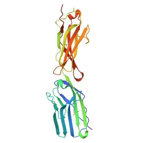
- Structurally, BCAM contains immunoglobulin domains, a signal peptide, a transmembrane helix, and disulfide bonds, and is classified as a blood group antigen (Lutheran system).
Experimental Guidance and Technical Tips
- The immunogen corresponds to amino acids 32-547 of human CD239/BCAM, encompassing the extracellular domain. This design makes the antibody well-suited for detecting surface CD239 on live cells by flow cytometry without permeabilization.
- Consider using appropriate isotype controls and titrating the antibody to determine the optimal staining concentration for your specific cell type.
- Given the broad tissue expression, validate antibody performance in samples where CD239 is expected to be highly expressed, such as human pancreatic or endothelial cells, and include a low-expression control (e.g., brain tissue-derived cells).
- As a monoclonal antibody, it offers consistent lot-to-lot reproducibility, which is advantageous for longitudinal studies or diagnostic assay development.
CamelBio: Your One-Stop Sourcing Bridge
CamelBio provides diagnostic manufacturers, labs, and research institutes with one-stop access to IVD raw materials, technical services, and consulting—covering every stage from concept to clinic. This Rabbit anti-Human CD239/BCAM monoclonal antibody exemplifies our commitment to supplying high-quality reagents for cell adhesion and oncology research. Our portfolio includes validated antibody pairs, optimized monoclonal and polyclonal antibodies, bulk ancillary reagents, and rare target sourcing to accelerate your assay development.
Product Datasheet
Rabbit anti-Human CD239/BCAM Monoclonal Antibody for Flow Cytometry - P50895
REQUEST A QUOTE
Our professional team will reply to you within one business day. Please feel free to contact us!