Monoclonal Antibodies
Rabbit anti-Human CD235a/Glycophorin A mAb for FC - P02724
Item Number : CM0008434
Price varies based on specs and customizations
- Application
- FC
- Cross Reactivity
- Human
- Protein Weight
- 13kDa/16kDa

Shipping:
Contact us to get shipping details Enjoy On-time Dispatch Guarantee.
Why Choose Us
Easy ordering process, quality products, and dedicated support for your business success.
Core Product Specifications and Parameters
| Parameter | Value |
|---|---|
| Product Name | Rabbit anti-Human CD235a/Glycophorin A mAb |
| Remarks/Alias | MN; GPA; MNS; GPSAT; PAS-2; CD235a; GPErik; HGpMiV; HGpMiXI; HGpSta(C) |
| Species | Human |
| GeneID (Human) | 2993 |
| GeneID | 2993 |
| Immunogen | Recombinant fusion protein containing a sequence corresponding to amino acids 20-91 of human CD235a (NP_002090.4). |
| Source | Rabbit |
| Category | Monoclonal Antibodies |
| Application | FC |
| Cross Reactivity | Human |
| SWISS | P02724 |
| Protein Weight | 13kDa/16kDa |
| Shipping | Ice bag |
Biological Background: Glycophorin A Function and Localization
- Glycophorin A (GYPA; aliases MN sialoglycoprotein, PAS-2, CD235a) is the major intrinsic membrane protein of human erythrocytes, encoded by the GYPA gene on chromosome 4.
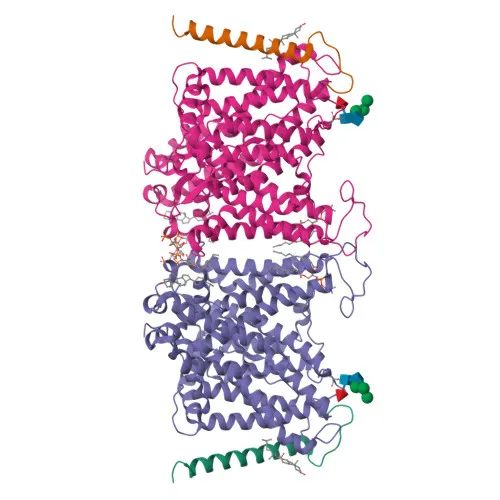
- It is a core component of the ankyrin-1 complex, which maintains erythrocyte membrane stability and shape. Disruption of this complex can lead to hereditary spherocytosis or elliptocytosis.
Related references:
PMID:35835865 - The extracellular N-terminal domain carries sialylated O-linked glycans that define the MN blood group system; the M and N antigens arise from amino acid polymorphisms at positions 1 and 5.
- Glycophorin A facilitates the trafficking and high-activity function of the anion exchanger SLC4A1 (band 3) to the plasma membrane.
- (Microbial infection) Acts as a receptor for Hepatitis A virus (HAV) and for Plasmodium falciparum erythrocyte-binding antigen 175 (EBA-175), with binding dependent on sialic acid residues.
- Subcellular localization is the plasma membrane, with a single transmembrane helix; the protein undergoes alternative splicing, phosphorylation, and O-linked glycosylation.
Experimental Guidance and Technical Tips
- The immunogen spans the N-terminal extracellular region (aa 20–91), which includes the MN antigenic site; therefore the antibody likely recognizes an epitope in this domain. Consider using red blood cell lysates or purified glycophorin A for positive controls.
- For flow cytometry, titrate the antibody to determine the optimal staining concentration; fix and permeabilize cells only if intracellular epitope access is required (though the target is extracellular).
- Validate the antibody in the appropriate sample system (e.g., human whole blood or erythrocyte-enriched fractions) and include an isotype control to assess non-specific binding.
- If studying malaria invasion or HAV attachment, perform functional blocking assays in parallel to confirm receptor interaction.
CamelBio: Your One-Stop Sourcing Bridge
CamelBio provides diagnostic manufacturers, labs, and research institutes with streamlined access to a broad portfolio of IVD raw materials, including validated antibody pairs, optimized monoclonal and polyclonal antibodies, bulk ancillary reagents, and rare target sourcing solutions. For researchers focusing on erythrocyte biology, blood group serology, or infectious disease mechanisms involving glycophorin A, CamelBio offers one-stop procurement support—from concept to clinic—helping you accelerate assay development and scale-up with confidence.
REQUEST A QUOTE
Our professional team will reply to you within one business day. Please feel free to contact us!