Monoclonal Antibodies
Rabbit anti-Human CD2 Monoclonal Antibody for Flow Cytometry - P06729
Item Number : CM0010833
Price varies based on specs and customizations
- Application
- FC
- Cross Reactivity
- Human
- Protein Weight
- 39kDa

Shipping:
Contact us to get shipping details Enjoy On-time Dispatch Guarantee.
Why Choose Us
Easy ordering process, quality products, and dedicated support for your business success.
Core Product Specifications and Parameters
| Parameter | Value |
|---|---|
| Product Name | Rabbit anti-Human CD2 mAb |
| Remarks/Alias | T11; SRBC; LFA-2 |
| Species | Human |
| Gene ID | 914 |
| Immunogen | Recombinant fusion protein containing a sequence corresponding to amino acids 25-209 of human CD2 (NP_001758.2) |
| Source | Rabbit |
| Category | Monoclonal Antibodies |
| Application | FC |
| Cross Reactivity | Human |
| SWISS | P06729 |
| Protein Weight | 39kDa |
| Shipping | Ice bag |
Biological Background: CD2 Function and Localization
- CD2 is a T-cell surface antigen, also known as LFA-2, SRBC receptor, and T11/Leu-5, belonging to the immunoglobulin superfamily. It mediates cell adhesion through interactions with its ligands.
- It interacts with lymphocyte function-associated antigen CD58 (LFA-3) and CD48/BCM1 to mediate adhesion between T-cells and other cell types, playing a critical role in T-cell activation.
- The cytoplasmic domain of CD2 is implicated in signal transduction, contributing to T-cell triggering and costimulatory functions.
- Expressed on T lymphocytes and natural killer cells; at the protein level, it is specifically detected in natural killer cells.
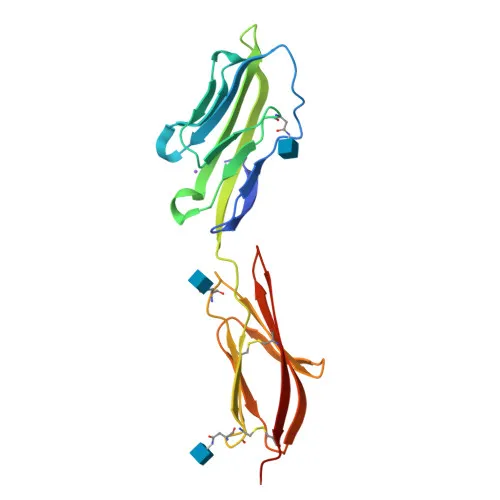
- CD2 is a single-pass type I membrane protein with two extracellular immunoglobulin-like domains, a transmembrane helix, and a cytoplasmic tail. The extracellular domain contains disulfide bonds and N-glycosylation sites.
- The CD2 gene is located on chromosome 1p13.1 and encodes a precursor with a signal peptide. Its molecular weight is approximately 39 kDa, consistent with the mature glycoprotein form.
- Keywords assigned to CD2 include cell adhesion, immunoglobulin domain, transmembrane, glycoprotein, disulfide bond, and signal, reflecting its structural and functional features.
Experimental Guidance and Technical Tips
- This antibody is designed for flow cytometry (FC) with human samples. For surface staining, use live unfixed cells to preserve the native conformation of the CD2 extracellular epitope.
- Consider titrating the antibody to determine the optimal staining concentration for your specific experimental setup, ensuring a high signal-to-noise ratio.
- Include appropriate isotype controls and fluorescence-minus-one (FMO) controls to accurately gate CD2-positive populations.
- If performing intracellular staining, validate the fixation and permeabilization protocol, as it may affect the recognition of the recombinant immunogen-derived epitope.
- For multicolor flow cytometry panels, check for potential spectral overlap with other fluorophores and perform compensation as needed.
CamelBio: Your One-Stop Sourcing Bridge
CamelBio offers diagnostic manufacturers and researchers a streamlined single-window for IVD raw-material procurement. This Rabbit anti-Human CD2 monoclonal antibody supports immunophenotyping and T-cell research, and we can supply additional validated antibody pairs, optimized clones, and bulk ancillary reagents. Our vertical integration ensures traceable, scalable sourcing from concept to clinic, covering rare-target antibody development and custom conjugation services.
REQUEST A QUOTE
Our professional team will reply to you within one business day. Please feel free to contact us!