Monoclonal Antibodies
Rabbit anti-Human CD1c Monoclonal Antibody for Flow Cytometry - P29017
Item Number : CM0005987
Price varies based on specs and customizations
- Application
- FC
- Cross Reactivity
- Human
- Protein Weight
- 38kDa

Shipping:
Contact us to get shipping details Enjoy On-time Dispatch Guarantee.
Why Choose Us
Easy ordering process, quality products, and dedicated support for your business success.
Core Product Specifications and Parameters
| Parameter | Value |
|---|---|
| Product Name | Rabbit anti-Human CD1c mAb |
| Remarks/Alias | R7; CD1; CD1A; BDCA1 |
| Species | Human |
| Gene ID (Human) | 911 |
| Gene ID | 911 |
| Immunogen | Recombinant fusion protein containing a sequence corresponding to amino acids 19-298 of human CD1c (NP_001756.2) |
| Source | Rabbit |
| Category | Monoclonal Antibodies |
| Application | FC |
| Cross Reactivity | Human |
| SWISS | P29017 |
| Protein Weight | 38kDa |
| Shipping | Ice bag |
Biological Background: CD1c Function and Localization
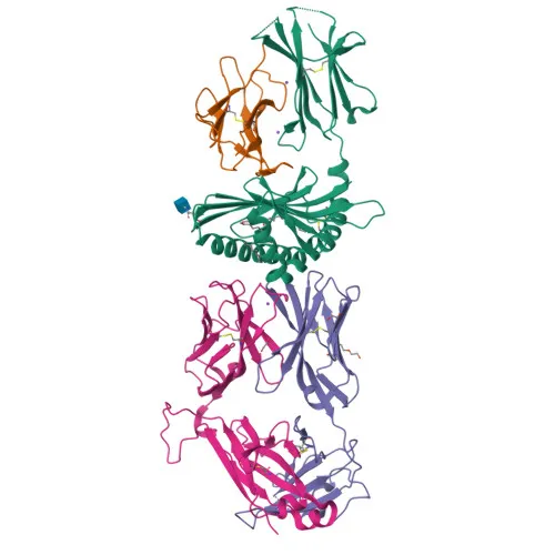
- CD1c is a member of the CD1 family of glycoproteins, which are structurally related to MHC class I molecules and mediate the presentation of lipid and glycolipid antigens to T cells.
- It functions as an antigen-presenting protein that binds self and non-self lipids and presents them to T-cell receptors on natural killer T-cells, playing a role in adaptive immunity.
- CD1c is expressed on the surface of professional antigen-presenting cells, including dendritic cells and B cells, and is known as a marker for the BDCA1+ subset of myeloid dendritic cells.
- The protein localizes to the cell membrane and to the membranes of endosomes and lysosomes, consistent with its role in capturing and presenting lipid antigens from intracellular compartments.
- According to UniProt, CD1c contains an immunoglobulin-like domain, a transmembrane helix, and a disulfide bond, and is subject to post-translational modifications such as glycosylation.
- It is also expressed on cortical thymocytes and certain T-cell leukemias, and has been detected in various other tissues.
Experimental Guidance and Technical Tips
- For flow cytometry, it is recommended to titrate the antibody to determine the optimal working concentration for your specific cell type and experimental conditions.
- Use appropriate isotype controls and viability dyes to ensure specific staining and exclude dead cells.
- Although the immunogen corresponds to the extracellular domain (aa 19-298), validation in the relevant human sample system (e.g., PBMCs or dendritic cell preparations) is advisable.
- As this is a directly conjugated antibody, avoid freeze-thaw cycles and protect from light to maintain fluorophore stability.
- Consult the manufacturer’s instructions for recommended buffer conditions and staining protocols.
CamelBio: Your One-Stop Sourcing Bridge
CamelBio serves as a one-stop sourcing bridge for IVD raw materials, offering validated antibody pairs, bulk ancillary reagents, and rare target raw-material sourcing to diagnostic manufacturers and research institutes. Our portfolio includes optimized monoclonal and polyclonal antibodies for immunology targets like CD1c, supporting applications from flow cytometry to immunohistochemistry. Contact our team to explore how we can streamline your supply chain from concept to clinic.
Product Datasheet
Rabbit anti-Human CD1c Monoclonal Antibody for Flow Cytometry - P29017
REQUEST A QUOTE
Our professional team will reply to you within one business day. Please feel free to contact us!