Monoclonal Antibodies
Rabbit anti-Human CD197/CCR7 Monoclonal Antibody for Flow Cytometry - P32248
Item Number : CM0007918
Price varies based on specs and customizations
- Application
- FC
- Cross Reactivity
- Human
- Protein Weight
- 43kDa

Shipping:
Contact us to get shipping details Enjoy On-time Dispatch Guarantee.
Why Choose Us
Easy ordering process, quality products, and dedicated support for your business success.
Core Product Specifications and Parameters
| Parameter | Value |
|---|---|
| Product Name | Rabbit anti-Human CD197/CCR7 mAb |
| Remarks/Alias | BLR2; EBI1; CCR-7; CD197; CDw197; CMKBR7; CC-CKR-7 |
| Species | Human |
| Gene ID | 1236 |
| Immunogen | Recombinant fusion protein containing a sequence corresponding to amino acids 25-59 of human CD197/CCR7 (NP_001829.1) |
| Source | Rabbit |
| Category | Monoclonal Antibodies |
| Application | FC |
| Cross Reactivity | Human |
| SWISS | P32248 |
| Protein Weight | 43kDa |
| Shipping | Ice bag |
Biological Background: CD197/CCR7 Function and Localization
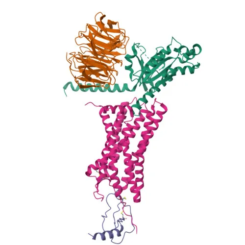
- CD197/CCR7 (C-C chemokine receptor type 7) is also known as BLR2, EBI1, CDw197, and MIP-3 beta receptor. It belongs to the G protein-coupled receptor (GPCR) family and mediates chemotactic lymphocyte migration.
- It functions as the receptor for the MIP-3-beta chemokine (CCL19) and is implicated in mediating Epstein-Barr virus (EBV) effects on B-lymphocytes and normal lymphocyte functions.
- CCR7 is a multi-pass transmembrane protein localized at the cell membrane, where it transduces extracellular signals via G proteins.
- Expression is primarily in lymphoid tissues and on activated B- and T-lymphocytes. It is strongly upregulated in B-cells infected with EBV and T-cells infected with herpesvirus 6 or 7.
- The protein contains key structural features typical of chemokine receptors: an extracellular N-terminal domain, seven transmembrane helices, disulfide bonds, and N-linked glycosylation sites.
- UniProt keywords include: 3D-structure, cell membrane, disulfide bond, G protein-coupled receptor, glycoprotein, membrane, receptor, signal, transducer, and transmembrane helix.
Experimental Guidance and Technical Tips
- This antibody is designed for flow cytometry (FC) and is conjugated to for direct detection. Verify fluorescence compatibility with your instrument's channels.
- The immunogen comprises the extracellular N-terminal region (amino acids 25-59), which is exposed on the cell surface; thus, the antibody is expected to recognize native membrane-expressed CCR7 without permeabilization.
- Perform titration experiments to determine the optimal antibody concentration for your specific cell type and experimental conditions. Use an appropriate isotype control to assess non-specific binding.
- Include positive control cells such as activated human peripheral blood lymphocytes or CCR7-transfected cell lines. Keep samples protected from light after staining to prevent fluorophore photobleaching.
- As CCR7 expression is upregulated by certain herpesviruses, consider validating in EBV- or HHV-6/7-infected lymphocyte models if relevant.
CamelBio: Your One-Stop Sourcing Bridge
CamelBio supports diagnostic manufacturers and research institutions with comprehensive access to IVD raw materials, including validated antibody pairs, bulk monoclonal antibodies, and ancillary reagents. For studies of lymphocyte trafficking, immune cell profiling, or viral pathology, this CCR7 antibody can be integrated into your flow cytometry panels. CamelBio's one-stop sourcing capabilities simplify procurement from concept to clinical application, ensuring consistent quality and supply for your development needs.
Product Datasheet
Rabbit anti-Human CD197/CCR7 Monoclonal Antibody for Flow Cytometry - P32248
REQUEST A QUOTE
Our professional team will reply to you within one business day. Please feel free to contact us!