Monoclonal Antibodies
Rabbit anti-Human CD120b/TNFRSF1B mAb for FC - P20333
Item Number : CM0006095
Price varies based on specs and customizations
- Application
- FC
- Cross Reactivity
- Human
- Protein Weight
- 28kDa/48kDa

Shipping:
Contact us to get shipping details Enjoy On-time Dispatch Guarantee.
Why Choose Us
Easy ordering process, quality products, and dedicated support for your business success.
Core Product Specifications and Parameters
| Parameter | Value |
|---|---|
| Product Name | Rabbit anti-Human CD120b/TNFRSF1B mAb |
| Remarks/Alias | p75; TBPII; TNFBR; TNFR2; CD120b; TNFR1B; TNFR80; TNF-R75; p75TNFR; TNF-R-II |
| Species | Human |
| GeneID (human) | 7133 |
| GeneID | 7133 |
| Immunogen | Recombinant fusion protein containing a sequence corresponding to amino acids 23-257 of human CD120b/TNFRSF1B (NP_001057.1) |
| Source | Rabbit |
| Category | Monoclonal Antibodies |
| Application | FC |
| Cross Reactivity | Human |
| SWISS | P20333 |
| Protein Weight | 28kDa/48kDa |
| Shipping | Ice bag |
Biological Background: TNFRSF1B Function and Localization
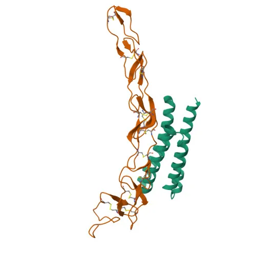
- TNFRSF1B (also known as TNFR2, p75, CD120b) is a member of the tumor necrosis factor receptor superfamily. It is a high-affinity receptor for TNF and has lower affinity for lymphotoxin-alpha.
- The receptor mediates most of the metabolic effects of TNF and plays a role in apoptosis regulation through the recruitment of TRAF1/TRAF2 complex, which then recruits apoptotic suppressors BIRC2 and BIRC3.
- Alternatively spliced isoform 2 lacking the transmembrane domain is secreted and blocks TNF-induced apoptosis, suggesting a regulatory function by antagonizing TNF biological activity.
- Localized primarily to the cell membrane as a single-pass type I membrane protein; a secreted form exists due to alternative splicing or proteolytic cleavage.
- Contains disulfide bonds and is extensively glycosylated, contributing to observed protein weights (e.g., ~48 kDa predicted, with additional bands due to modifications). It is also phosphorylated.
- The protein has implications in pharmaceutical research, being a target for anti-inflammatory therapies.
Experimental Guidance and Technical Tips
- The immunogen corresponds to the extracellular domain (23-257), suggesting the antibody targets the extracellular region, making it suitable for live-cell flow cytometry without permeabilization.
- For best results in flow cytometry, titrate the antibody to determine the optimal concentration for your specific cell type and experimental conditions.
- Include appropriate isotype controls and viability dyes to distinguish specific staining from background and dead cells.
- Given the potential shedding of TNFR2, consider analyzing both surface and soluble receptor forms if studying regulation.
- Since the antibody is directly conjugated to , it enables one-step staining, reducing assay time and potential variability from secondary reagents.
CamelBio: Your One-Stop Sourcing Bridge
CamelBio provides diagnostic manufacturers and research laboratories with a streamlined supply chain for IVD raw materials, including validated antibody pairs, monoclonal and polyclonal antibodies, and ancillary reagents. This TNFRSF1B/TNFR2 antibody supports apoptosis and inflammatory disease research, and CamelBio can source additional rare targets or bulk reagents upon request.
REQUEST A QUOTE
Our professional team will reply to you within one business day. Please feel free to contact us!