Polyclonal Antibodies
Rabbit Anti-HPCAL1 Polyclonal Antibody for WB and ELISA - P37235
Item Number : CM0024317
Price varies based on specs and customizations
- Application
- WB, ELISA
- Cross Reactivity
- Human, Mouse, Rat
- Protein Weight
- 22kDa

Shipping:
Contact us to get shipping details Enjoy On-time Dispatch Guarantee.
Why Choose Us
Easy ordering process, quality products, and dedicated support for your business success.
Core Product Specifications and Parameters
| Parameter | Value |
|---|---|
| Product Name | [KO Validated] HPCAL1 Rabbit pAb |
| Remarks/Alias | BDR1; HLP2; VILIP-3; L1 |
| Species | Human |
| GeneID (Human) | 3241 |
| GeneID | 3241 |
| Immunogen | Recombinant fusion protein containing a sequence corresponding to amino acids 1-193 of human HPCAL1 (NP_002140.2). |
| Source | Rabbit |
| Category | Polyclonal Antibodies |
| Application | WB, ELISA |
| Cross Reactivity | Human, Mouse, Rat |
| SWISS | P37235 |
| Protein Weight | 22kDa |
| Shipping | Ice bag |
Biological Background: HPCAL1 Function and Localization
- HPCAL1, also known as Hippocalcin-like protein 1, calcium-binding protein BDR-1, HLP2, or Visinin-like protein 3 (VILIP-3), belongs to the neuronal calcium sensor (NCS) family of EF-hand calcium-binding proteins.
- The protein contains multiple functional EF-hand motifs and an N-terminal myristoylation consensus sequence, characteristic of calcium-dependent membrane-associating proteins (Keywords: Calcium, Lipoprotein, Myristate, Repeat).
- HPCAL1 may be involved in the calcium-dependent regulation of rhodopsin phosphorylation, implicating it in phototransduction or related G-protein coupled receptor signaling pathways.
- Subcellularly, HPCAL1 is membrane-associated, likely mediated by its N-terminal myristoyl group in a calcium-dependent manner (SUBCELLULAR LOCATION: Membrane).
- Post-translational N-myristoylation is essential for its proper membrane targeting and function, as indicated by the Myristate keyword.
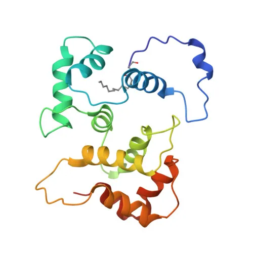
- The protein has been structurally characterized (3D-structure keyword), revealing calcium-induced conformational changes typical of NCS proteins.
- HPCAL1 is included in the human reference proteome and has been identified through large-scale proteomics studies (Proteomics identification keyword).
Experimental Guidance and Technical Tips
- The immunogen encompasses the full-length human HPCAL1 protein (residues 1-193), fused to a recombinant tag. For Western blotting, validate specificity by comparing signals between HPCAL1 knockout and wild-type cell or tissue lysates.
- In WB, expect a band at approximately 22 kDa. Always include appropriate molecular weight markers and loading controls to confirm protein identity.
- For ELISA, optimize coating antigen and antibody concentrations through titration. If cross-reactivity with related visinin-like proteins is a concern, consider pre-adsorption or competitive assays.
- Although this antibody is KO-validated by the supplier, we recommend independent validation in your specific experimental system to confirm performance and suitability.
CamelBio: Your One-Stop Sourcing Bridge
CamelBio serves as a comprehensive sourcing partner for IVD raw materials, offering diagnostic manufacturers and research labs reliable antibodies, antigens, and ancillary reagents. This anti-HPCAL1 polyclonal antibody supports neuroscience and calcium signaling research, with applications in WB and ELISA. Beyond catalog products, we provide custom antibody development, bulk supply, and rare-target sourcing to streamline your entire development pipeline from concept to clinic.
REQUEST A QUOTE
Our professional team will reply to you within one business day. Please feel free to contact us!