Polyclonal Antibodies
PPARα Rabbit pAb - Q07869
Item Number : CM0028140
Price varies based on specs and customizations
- Application
- WB, ELISA
- Cross Reactivity
- Mouse, Rat
- Protein Weight
- 52kDa

Shipping:
Contact us to get shipping details Enjoy On-time Dispatch Guarantee.
Why Choose Us
Easy ordering process, quality products, and dedicated support for your business success.
Core Product Specifications and Parameters
| Parameter | Value |
|---|---|
| Product Name | PPARα Rabbit pAb |
| Remarks/Alias | PPAR; NR1C1; hPPAR; PPARalpha; PPAR-alpha; PPARα |
| Species | Human |
| GeneID (Human) | 5465 |
| GeneID | 5465 |
| Immunogen | Recombinant fusion protein containing a sequence corresponding to amino acids 1-92 of human PPARα (NP_005027.2). |
| Source | Rabbit |
| Category | Polyclonal Antibodies |
| Application | WB, ELISA |
| Cross Reactivity | Mouse, Rat |
| SWISS | Q07869 |
| Protein Weight | 52kDa |
| Shipping | Ice bag |
Biological Background: PPARα Function and Localization
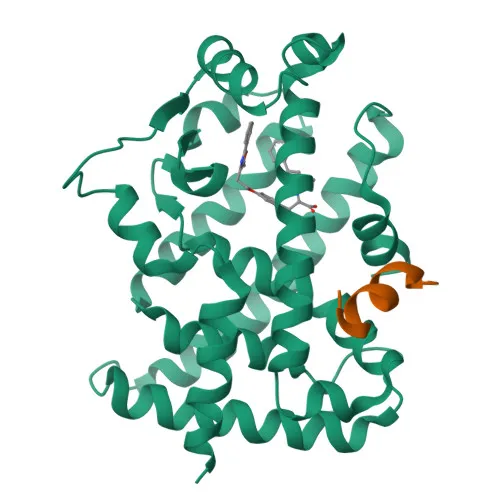
- PPARα (Peroxisome proliferator-activated receptor alpha), encoded by the PPARA gene (also known as NR1C1), is a member of the nuclear receptor superfamily of transcription factors.
- It functions as a ligand-activated transcription factor and is a master regulator of lipid metabolism, controlling fatty acid oxidation and energy homeostasis.
- PPARα is activated by endogenous ligands such as 1-palmitoyl-2-oleoyl-sn-glycerol-3-phosphocholine (16:0/18:1-GPC) and oleylethanolamide, as well as by synthetic peroxisome proliferators including hypolipidemic drugs.
- Upon activation, PPARα upregulates the expression of genes involved in the peroxisomal beta-oxidation pathway, including ACOX1 and cytochrome P450 enzymes.
- Transcriptional activity of PPARα requires heterodimerization with retinoid X receptor alpha (RXRA) and is antagonized by the nuclear receptor NR2C2.
- PPARα has been implicated in the circadian clock system, where it may propagate timing signals from PER2 to metabolic pathways, linking biological rhythms with lipid metabolism.
- It is predominantly expressed in metabolically active tissues, including skeletal muscle, liver, heart, and kidney, and has been detected in monocytes (PubMed:28167758). Related references: PMID:28167758
- Subcellular localization is primarily nuclear, consistent with its role as a DNA-binding transcription factor.
- Structural features include zinc-finger DNA-binding domains, and the protein is subject to post-translational modifications such as ubiquitin-like conjugation.
Experimental Guidance and Technical Tips
- The immunogen corresponds to amino acids 1-92 at the N-terminus of human PPARα, in a region that may be less conserved among nuclear receptors; consider validating specificity for PPARα in your target species.
- For Western blotting, the predicted molecular weight is approximately 52 kDa. Verify band detection in your lysate system using appropriate controls, and note the antibody's cross-reactivity with mouse and rat samples.
- In ELISA, the antibody may be suitable for detecting recombinant or endogenous PPARα; optimize coating conditions and dilution factors for your assay format.
- Polyclonal antibodies may exhibit lot-to-lot variability; we recommend performing lot-specific validation for critical quantitative applications.
CamelBio: Your One-Stop Sourcing Bridge
As a one-stop IVD raw-material sourcing partner, CamelBio supports diagnostic manufacturers and research labs with high-quality antibodies, validated pairs, and ancillary reagents tailored for lipid metabolism and nuclear receptor studies. This PPARα polyclonal antibody is part of our extensive portfolio for metabolic disease biomarker development. Beyond standard catalog products, we offer custom monoclonal antibody development, bulk production, and rare-target sourcing to accelerate your assay development from concept to clinic.
REQUEST A QUOTE
Our professional team will reply to you within one business day. Please feel free to contact us!