Monoclonal Antibodies
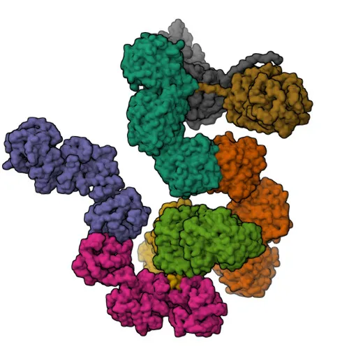
PLOD3 Rabbit mAb - O60568
Item Number : CM0002894
Price varies based on specs and customizations
- Application
- WB, IHC-P, ELISA
- Cross Reactivity
- Human
- Protein Weight
- 85kDa

Shipping:
Contact us to get shipping details Enjoy On-time Dispatch Guarantee.
Why Choose Us
Easy ordering process, quality products, and dedicated support for your business success.
Core Product Specifications and Parameters
| Parameter | Value |
|---|---|
| Product Name | PLOD3 Rabbit mAb |
| Remarks/Alias | LH3; PLOD3 |
| Species | Human |
| Gene ID (Human) | 8985 |
| Gene ID | 8985 |
| Immunogen | Recombinant protein / Recombinant fusion protein containing a sequence corresponding to amino acids 25-306 of human PLOD3 (NP_001075.1). |
| Source | Rabbit |
| Category | Monoclonal Antibodies |
| Application | WB, IHC-P, ELISA |
| Cross Reactivity | Human |
| SWISS | O60568 |
| Protein Weight | 85kDa |
| Shipping | Ice bag |
Biological Background: PLOD3 Function and Localization
- Protein identity and function: PLOD3, also known as LH3 (lysyl hydroxylase 3), is a multifunctional enzyme that performs critical post-translational modifications on collagen lysine residues.
- Hydroxylase activity: It catalyzes the hydroxylation of lysine in -Xaa-Lys-Gly- motifs of procollagens, generating hydroxylysine. This step is essential for collagen thermal stability and cross-link formation. Related references: PMID:11956192, PMID:12475640, PMID:18298658
- Glycosyltransferase activities: Following hydroxylation, the enzyme sequentially adds galactose and glucose to hydroxylysine, forming 1,2-glucosylgalactosyl-5-hydroxylysine. These glycosylation events are indispensable for type IV collagen secretion, basement membrane assembly, and also occur on MBL1 collagen-like domain. Related references: PMID:12475640, PMID:18298658, PMID:18834968, PMID:10934207, PMID:11896059, PMID:11956192, PMID:25419660
- Subcellular localization: PLOD3 resides predominantly in the rough endoplasmic reticulum (ER) lumen and membrane, with a minor pool secreted into the extracellular space. This is consistent with its role in collagen processing within the secretory pathway.
- Tissue distribution: Ubiquitously expressed, with highest levels in heart, placenta, and pancreas, and lower levels in lung, liver, and skeletal muscle. Related references: PMID:9582318, PMID:9724729
- Cofactor requirements: The hydroxylase domain depends on Fe²⁺, 2-oxoglutarate, O₂, and ascorbate (vitamin C), while the glycosyltransferase activities likely require Mn²⁺.
- Structural features: The enzyme is itself a glycoprotein with intramolecular disulfide bonds.
Experimental Guidance and Technical Tips
- Western blotting: Under reducing and denaturing conditions, a specific band at approximately 85 kDa should be observed. Use appropriate protein loading and validate with a relevant cell line or tissue lysate expressing PLOD3.
- Immunohistochemistry (paraffin): Standard antigen retrieval (e.g., citrate buffer, pH 6.0) and blocking steps are recommended. Optimization of primary antibody concentration and detection system is advised.
- ELISA: The antibody can be used in direct or capture ELISA formats. Titration of both coating antigen and antibody concentration should be performed to determine optimal conditions.
- Immunogen considerations: This antibody was raised against a recombinant protein spanning amino acids 25–306 (N-terminal region). It may detect both full-length PLOD3 and potential N-terminal fragments.
CamelBio: Your One-Stop Sourcing Bridge
CamelBio is a trusted one-stop supplier of IVD raw materials, connecting diagnostic developers with high-quality reagents like this anti-PLOD3 monoclonal antibody for collagen modification research. We offer validated antibody pairs, optimized recombinant proteins, bulk ancillary reagents, and sourcing of rare targets to accelerate your assay development from concept to clinic.
REQUEST A QUOTE
Our professional team will reply to you within one business day. Please feel free to contact us!