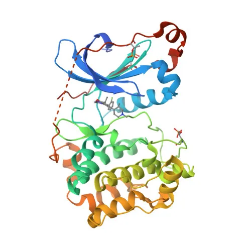
Monoclonal Antibodies
PKC alpha Rabbit mAb for IF/ICC, ELISA - P17252
Item Number : CM0007536
Price varies based on specs and customizations
- Application
- IF/ICC, ELISA
- Cross Reactivity
- Human
- Protein Weight
- 77kDa

Shipping:
Contact us to get shipping details Enjoy On-time Dispatch Guarantee.
Why Choose Us
Easy ordering process, quality products, and dedicated support for your business success.
Core Product Specifications and Parameters
| Parameter | Value |
|---|---|
| Product Name | PKC alpha Rabbit mAb |
| Remarks/Alias | AAG6; PKCA; PRKACA; PKCI+/-; PKCalpha; PKC-alpha |
| Species | Human |
| GeneID | 5578 |
| Immunogen | Recombinant protein of human PKC alpha |
| Source | Rabbit |
| Category | Monoclonal Antibodies |
| Application | IF/ICC, ELISA |
| Cross Reactivity | Human |
| SWISS | P17252 |
| Protein Weight | 77kDa |
| Shipping | Ice bag |
Biological Background: Protein kinase C alpha type Function and Localization
- Protein kinase C alpha type (PKCα), encoded by the PRKCA gene (aliases: PKCA, PRKACA), is a calcium-activated, phospholipid- and diacylglycerol (DAG)-dependent serine/threonine-protein kinase. It belongs to the PKC family of kinases, which play key roles in diverse signal transduction pathways.
- PKCα is involved in the positive and negative regulation of cell proliferation, apoptosis, differentiation, migration, adhesion, tumorigenesis, cardiac hypertrophy, angiogenesis, platelet function, and inflammation. It directly phosphorylates targets like RAF1, BCL2, CSPG4, TNNT2/CTNT, and activates MAPK/ERK and RAP1GAP signaling cascades.
- In cancer biology, PKCα is highly expressed in various tumors, including gliomas and breast cancers, where it can act as a tumor promoter. It exhibits anti-apoptotic functions by suppressing p53-mediated IGFBP3 activation and phosphorylating BCL2. In glioma cells, it promotes cell growth via RAF1/MAPK/ERK.
- PKCα regulates cytoskeletal dynamics and cell migration. For example, it phosphorylates CSPG4, inducing lamellipodia formation and cell polarization. During chemokine-induced CD4+ T cell migration, PKCα phosphorylates DOCK8, leading to CDC42 activation and enhanced motility. Related references: PMID:28028151
- The subcellular localization of PKCα is dynamic: it shuttles between the cytoplasm, cell membrane, mitochondrial membrane, and nucleus, depending on stimulation. This trafficking is critical for its signaling functions in processes like macrophage differentiation and wound healing.
- In the cardiovascular system, PKCα negatively regulates myocardial contractility by phosphorylating cardiac troponin T (TNNT2/CTNT), which reduces actomyosin ATPase activity and calcium sensitivity. It also promotes cardiac hypertrophy through MAPK/ERK signaling and is essential for angiogenesis, mediating endothelial cell migration and VEGFA-dependent signaling.
- PKCα is subject to post-translational modifications, including phosphorylation and acetylation, which modulate its activity and interactions. It is also a proto-oncogene and a reference proteome entry (UniProt keywords: Acetylation, Apoptosis, ATP-binding, Calcium, Cell adhesion, Cell membrane, Cytoplasm, Direct protein sequencing, Kinase, Membrane, Metal-binding, Mitochondrion, Nucleotide-binding, Nucleus, Phosphoprotein, Proteomics identification, Proto-oncogene, Reference proteome, Repeat, Serine/threonine-protein kinase, Transferase, Zinc, Zinc-finger).
- PKCα phosphorylates KLHL3 in response to angiotensin II, impacting WNK4 signaling and ion homeostasis. It also activates LRRK1, which then phosphorylates RAB proteins to regulate intracellular trafficking. Related references: PMID:25313067 PMID:36040231
Experimental Guidance and Technical Tips
- The immunogen used is recombinant human PKC alpha protein. This design typically yields antibodies recognizing native protein conformations. For IF/ICC, consider optimizing fixation/permeabilization conditions to preserve the target's subcellular localization (cytoplasm, membrane, nucleus).
- For ELISA, the antibody can be used to detect PKCα in cell lysates or purified protein samples. Pair it with a suitable detection system and validate in the relevant sample system to ensure specificity.
- PKCα activation often involves translocation; therefore, in IF/ICC experiments, stimulation with agonists like PMA may reveal dynamic redistribution patterns. Always include appropriate controls.
- Cross-reactivity is reported for human; verify reactivity with other species if needed.
CamelBio: Your One-Stop Sourcing Bridge
CamelBio provides diagnostic manufacturers, laboratories, and research institutes with one-stop access to IVD raw materials, technical services, and consulting. This PKC alpha monoclonal antibody is part of our extensive portfolio of validated antibody reagents for cell signaling and cancer research applications. Whether you need optimized antibody pairs, monoclonal/polyclonal antibodies, bulk ancillary reagents, or rare target raw-material sourcing, CamelBio supports your development from concept to clinic.
REQUEST A QUOTE
Our professional team will reply to you within one business day. Please feel free to contact us!