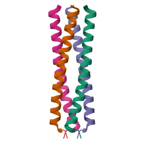
Phosphorylated Antibodies
Phospho-VASP-S239 Rabbit Monoclonal Antibody for WB, IF/ICC, ELISA - P50552
Item Number : CM0001906
Price varies based on specs and customizations
- Application
- WB, IF/ICC, ELISA
- Cross Reactivity
- Human
- Protein Weight
- 40kDa

Shipping:
Contact us to get shipping details Enjoy On-time Dispatch Guarantee.
Why Choose Us
Easy ordering process, quality products, and dedicated support for your business success.
Core Product Specifications and Parameters
| Parameter | Value |
|---|---|
| Product Name | Phospho-VASP-S239 Rabbit mAb |
| Remarks/Alias | VASP; vasodilator-stimulated phosphoprotein; Phospho-VASP-S239 |
| Species | Human |
| GeneID | 7408 |
| Immunogen | Synthetic peptide (A synthetic phosphorylated peptide around S239 of human VASP (NP_003361.1).) |
| Source | Rabbit |
| Category | Phosphorylated Antibodies |
| Application | WB, IF/ICC, ELISA |
| Cross Reactivity | Human |
| SWISS | P50552 |
| Protein Weight | 40kDa |
| Shipping | Ice bag |
Biological Background: Vasodilator-stimulated phosphoprotein (VASP) Function and Localization
- VASP is a member of the Ena/VASP protein family, which are actin-associated proteins involved in cytoskeleton remodeling and cell polarity.
- It promotes actin filament elongation by protecting filament barbed ends from capping proteins and enhancing the rate of actin polymerization.
- VASP facilitates the transfer of profilin-bound actin monomers to growing filament barbed ends, thereby stimulating actin filament elongation.
- It plays a crucial role in platelet activation and aggregation, regulating actin dynamics essential for platelet function.
- The protein is localized to various subcellular structures including the cytoplasm, focal adhesions, tight junctions, and the membrane of lamellipodia and filopodia.
- VASP is highly expressed in platelets, reflecting its specialized role in these cells.
- Phosphorylation at serine 239, among other sites, represents a key regulatory modification influencing VASP activity and interactions.
Experimental Guidance and Technical Tips
- The immunogen is a synthetic phosphorylated peptide around S239; thus, the antibody specifically detects VASP phosphorylated at this site. Include phosphatase inhibitors in lysis buffers to preserve phosphorylation status during sample preparation.
- For Western blot, platelet lysates are recommended due to high VASP expression; expect a band around 40 kDa.
- In immunofluorescence/immunocytochemistry, co-staining with actin markers or focal adhesion proteins may help visualize VASP localization to lamellipodia, filopodia, or focal adhesions.
- Consider using appropriate positive and negative controls (e.g., phosphatase-treated samples) to confirm phospho-specificity.
CamelBio: Your One-Stop Sourcing Bridge
CamelBio serves as a comprehensive sourcing partner for IVD raw materials, offering validated antibody pairs, optimized monoclonal and polyclonal antibodies, and bulk ancillary reagents. For cytoskeleton dynamics and platelet activation research, CamelBio provides reliable access to phosphorylation-specific detection tools like anti-phospho-VASP antibodies. Our supply network also supports rare target raw material sourcing, helping diagnostic manufacturers and laboratories streamline procurement from concept to clinic.
Product Datasheet
Phospho-VASP-S239 Rabbit Monoclonal Antibody for WB, IF/ICC, ELISA - P50552
REQUEST A QUOTE
Our professional team will reply to you within one business day. Please feel free to contact us!