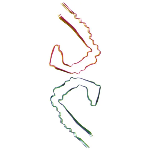
Phosphorylated Antibodies
Phospho-Tau-T231 Rabbit pAb - P10636
Item Number : CM0000701
Price varies based on specs and customizations
- Application
- WB, ELISA
- Cross Reactivity
- Mouse
- Protein Weight
- 79kDa

Shipping:
Contact us to get shipping details Enjoy On-time Dispatch Guarantee.
Why Choose Us
Easy ordering process, quality products, and dedicated support for your business success.
Core Product Specifications and Parameters
| Parameter | Value |
|---|---|
| Product Name | Phospho-Tau-T231 Rabbit pAb |
| Remarks/Alias | TAU; MSTD; PPND; DDPAC; MAPTL; MTBT1; MTBT2; tau-40; FTDP-17; PPP1R103; Tau-PHF6; Phospho-Tau-T231 |
| Species | Human |
| Gene ID | 4137 |
| Immunogen | Synthetic Peptide: A synthetic phosphorylated peptide around T548 of human Tau (NP_058519.3?) |
| Source | Rabbit |
| Category | Phosphorylated Antibodies |
| Application | WB, ELISA |
| Cross Reactivity | Mouse |
| SWISS | P10636 |
| Protein Weight | 79kDa |
| Shipping | Ice bag |
Biological Background: Tau Function and Localization
- Tau (microtubule-associated protein tau) is encoded by the MAPT gene and exists in multiple isoforms generated by alternative splicing, primarily expressed in neurons. The central nervous system forms differ from the peripheral nervous system isoform (PNS-tau).
- The core function of Tau is to promote microtubule assembly and stability, which is critical for establishing and maintaining neuronal polarity. Related references: PMID:21985311
- Tau acts as a linker protein by binding to axonal microtubules via its C-terminus and to neural plasma membrane components via its N-terminus. Related references: PMID:21985311, PMID:32961270
- Subcellularly, Tau is found in the cytoplasm, cytosol, associated with the cytoskeleton, and localized to axons and dendrites. It can also be secreted and associate with the cell membrane.
- Post-translational modifications of Tau include phosphorylation, acetylation, glycation, methylation, and ubiquitination. Hyperphosphorylation at sites like T231 is a hallmark of Alzheimer's disease and related tauopathies, leading to paired helical filament (PHF) formation.
- Tissue specificity: Isoforms are differentially expressed; CNS isoforms are found in the central nervous system, while PNS-tau is expressed in the peripheral nervous system.
Experimental Guidance and Technical Tips
- The immunogen was a synthetic phosphorylated peptide around T548 of human Tau (corresponding to T231 in the commonly studied 441-amino acid isoform), indicating specificity for the T231 phosphorylation site. Confirm the exact numbering in your target isoform.
- For Western blotting, use phospho‑specific lysis buffers and consider including phosphatase inhibitors to preserve phosphorylation. Verify band specificity with non‑phosphorylated Tau controls or phosphatase treatment.
- The antibody shows cross‑reactivity with mouse; however, validate the recognition of phosphorylated Tau at T231 in mouse samples, as phosphorylation patterns may differ between species.
- For ELISA applications, pairing with a capture antibody that recognizes total Tau may help to normalize phosphorylation signals.
CamelBio: Your One-Stop Sourcing Bridge
CamelBio provides diagnostic manufacturers and research laboratories with streamlined access to validated IVD raw materials, including phosphorylation‑specific antibodies such as this anti‑Phospho‑Tau‑T231 reagent. Beyond individual antibodies, CamelBio offers complementary reagents, bulk raw materials, and sourcing support to accelerate neurodegenerative disease biomarker assay development from concept to clinic.
REQUEST A QUOTE
Our professional team will reply to you within one business day. Please feel free to contact us!