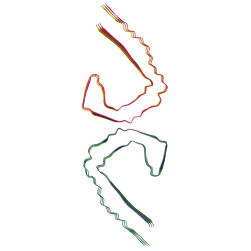
Phosphorylated Antibodies
Phospho-Tau-T181 Rabbit mAb for WB, ELISA - P10636
Item Number : CM0001891
Price varies based on specs and customizations
- Application
- WB, ELISA
- Cross Reactivity
- Human
- Protein Weight
- 79kDa

Shipping:
Contact us to get shipping details Enjoy On-time Dispatch Guarantee.
Why Choose Us
Easy ordering process, quality products, and dedicated support for your business success.
Core Product Specifications and Parameters
| Parameter | Value |
|---|---|
| Product Name | Phospho-Tau-T181 Rabbit mAb |
| Remarks/Alias | TAU; MSTD; PPND; DDPAC; MAPTL; MTBT1; MTBT2; tau-40; FTDP-17; PPP1R103; Tau-PHF6; Phospho-Tau-T181 |
| Species | Human |
| GeneID (Human) | 4137 |
| GeneID | 4137 |
| Immunogen | Synthetic Peptide|A synthetic phosphorylated peptide around T181 of human Phospho-Tau-T181 (NP_001190181.1). |
| Source | Rabbit |
| Category | Phosphorylated Antibodies |
| Application | WB, ELISA |
| Cross Reactivity | Human |
| SWISS | P10636 |
| Protein Weight | 79kDa |
| Shipping | Ice bag |
Biological Background: Tau Function and Localization
- Microtubule-associated protein tau (MAPT) is primarily expressed in neurons and plays a critical role in promoting microtubule assembly and stability. Related references: PMID:21985311, PMID:32961270
- Tau functions as a linker protein: its C-terminus binds axonal microtubules while its N-terminus associates with neural plasma membrane components, contributing to neuronal polarity. Related references: PMID:21985311, PMID:32961270
- The MAPT gene encodes multiple isoforms via alternative splicing; shorter isoforms promote cytoskeletal plasticity, whereas longer isoforms likely stabilize microtubules.
- In neurons, tau localizes to the cytoplasm, cytoskeleton, axons, dendrites, and can be secreted; its subcellular distribution is critical for neuronal function.
- Tissue-specific expression includes central nervous system isoforms and a peripheral nervous system-specific isoform (PNS-tau).
- Post-translational modifications such as phosphorylation (e.g., at T181), acetylation, methylation, and ubiquitination regulate tau function and are implicated in neurodegenerative diseases.
- Aberrant hyperphosphorylation of tau leads to paired helical filament (PHF) formation, a hallmark of Alzheimer disease and other tauopathies.
Experimental Guidance and Technical Tips
- For Western blot, consider using human neuronal cell lysates or brain tissue extracts; validate phospho-specificity by treating samples with lambda phosphatase to confirm loss of signal at the expected molecular weight (~79 kDa).
- In ELISA applications, pair this capture/detection antibody with a non-phospho Tau antibody to quantify site-specific phosphorylation levels.
- As the antibody is validated for human Tau only, cross-reactivity with other species should be verified experimentally if needed.
CamelBio: Your One-Stop Sourcing Bridge
CamelBio is your comprehensive partner for IVD raw-material sourcing, offering validated antibody pairs, optimized monoclonal/polyclonal antibodies, and bulk ancillary reagents. For Tau-targeted neurodegenerative disease research, we provide tailored solutions from concept to clinic, leveraging our expertise to streamline your diagnostic development.
REQUEST A QUOTE
Our professional team will reply to you within one business day. Please feel free to contact us!