Phosphorylated Antibodies
Phospho-Tau (Ser198/Ser199/Ser202) Rabbit Polyclonal Antibody for WB and ELISA - P10636
Item Number : CM0001557
Price varies based on specs and customizations
- Application
- WB, ELISA
- Cross Reactivity
- Human, Mouse, Rat
- Protein Weight
- 79kDa

Shipping:
Contact us to get shipping details Enjoy On-time Dispatch Guarantee.
Why Choose Us
Easy ordering process, quality products, and dedicated support for your business success.
Core Product Specifications and Parameters
| Parameter | Value |
|---|---|
| Product Name | Phospho-Tau-Ser198/Ser199/Ser202 Rabbit pAb |
| Remarks/Alias | TAU; MSTD; PPND; DDPAC; MAPTL; MTBT1; MTBT2; tau-40; FTDP-17; PPP1R103; Tau-PHF6; Phospho-Tau-Ser198/Ser199/Ser202 |
| Species | Human |
| Gene ID (Human) | 4137 |
| Gene ID | 4137 |
| Immunogen | Synthetic peptide |
| Source | Rabbit |
| Category | Phosphorylated Antibodies |
| Application | WB, ELISA |
| Cross Reactivity | Human, Mouse, Rat |
| SWISS | P10636 |
| Protein Weight | 79kDa |
| Shipping | Ice bag |
Biological Background: Tau Function and Localization
- Tau (microtubule-associated protein tau) is encoded by the MAPT gene and exists as multiple isoforms via alternative splicing, with short isoforms promoting cytoskeletal plasticity and longer isoforms stabilizing microtubules in neurons.
- Tau promotes microtubule assembly and stability, acting as a linker protein between axonal microtubules (via its C-terminus) and neural plasma membrane components (via its N-terminus), which is essential for neuronal polarity (PubMed:21985311, PubMed:32961270). Related references: PMID:21985311 PMID:32961270
- Tau localization to the axon is determined by the centrosome, thereby contributing to the establishment and maintenance of neuronal polarity.
- Subcellularly, tau is found in the cytoplasm, cytosol, cytoskeleton, and cell membrane; it localizes to axons and dendrites, and can also be secreted.
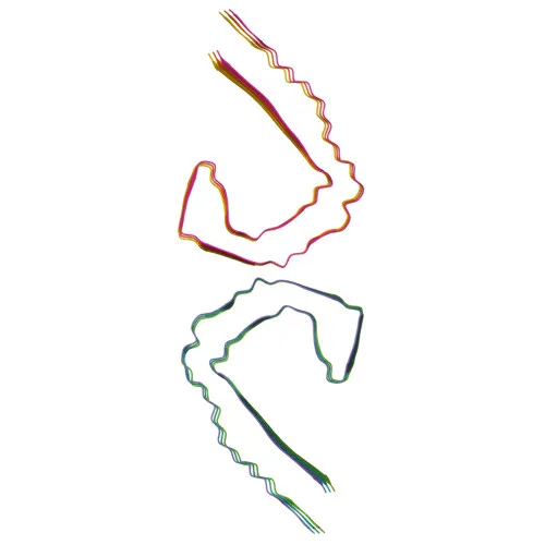
- Hyperphosphorylation of tau leads to its aggregation into paired helical filaments (PHF-tau), forming neurofibrillary tangles that are hallmark pathologies in Alzheimer disease and related tauopathies.
- Tau is predominantly expressed in neurons, with the PNS-tau isoform expressed in the peripheral nervous system and other isoforms in the central nervous system.
Experimental Guidance and Technical Tips
- The immunogen is a synthetic peptide phosphorylated at Ser198, Ser199, and Ser202; therefore, this antibody is expected to recognize Tau only when these residues are phosphorylated. Consider using phosphatase inhibitors during sample preparation to preserve phosphorylation.
- For Western blot, the expected molecular weight is approximately 79 kDa for the full-length isoform, but multiple bands may appear due to alternative splicing and phosphorylation states. Validate in relevant neuronal cell lines or brain tissue lysates.
- In ELISA, use phosphorylated peptide or protein standards to confirm specificity for the phosphorylated epitope.
- Cross-reactivity with mouse and rat has been reported; however, sequence alignment of the phospho-site should be verified for your specific target species and isoform.
CamelBio: Your One-Stop Sourcing Bridge
CamelBio supports diagnostic manufacturers and research laboratories with comprehensive access to IVD raw materials, including this anti-phospho-Tau antibody for neurodegeneration research and assay development. Our supply network offers validated antibody pairs, bulk ancillary reagents, and sourcing for rare targets, streamlining your path from concept to clinic.
Product Datasheet
Phospho-Tau (Ser198/Ser199/Ser202) Rabbit Polyclonal Antibody for WB and ELISA - P10636
REQUEST A QUOTE
Our professional team will reply to you within one business day. Please feel free to contact us!