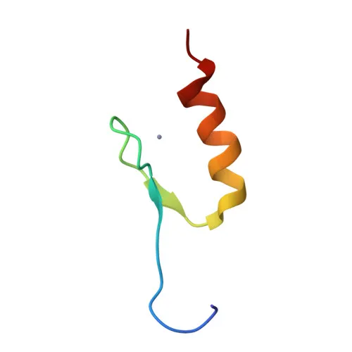
Phosphorylated Antibodies
Phospho-SP1 (T739) Rabbit Polyclonal Antibody for Western Blotting - P08047
Item Number : CM0000921
Price varies based on specs and customizations
- Application
- WB
- Cross Reactivity
- Human, Mouse, Rat
- Protein Weight
- 81kDa

Shipping:
Contact us to get shipping details Enjoy On-time Dispatch Guarantee.
Why Choose Us
Easy ordering process, quality products, and dedicated support for your business success.
Core Product Specifications and Parameters
| Parameter | Value |
|---|---|
| Product Name | Phospho-SP1-T739 Rabbit pAb |
| Remarks/Alias | SP1; Phospho-SP1-T739 |
| Species | Human |
| Gene ID | 6667 |
| Immunogen | A phospho specific peptide corresponding to residues surrounding T739 of human SP1 |
| Source | Rabbit |
| Category | Phosphorylated Antibodies |
| Application | WB |
| Cross Reactivity | Human, Mouse, Rat |
| SWISS | P08047 |
| Protein Weight | 81kDa |
| Shipping | Ice bag |
Biological Background: SP1 Function and Localization
- SP1 is a transcription factor that binds GC-rich promoter motifs to regulate genes involved in cell growth, apoptosis, differentiation, and immune responses.
- It is subject to extensive post-translational modifications, including phosphorylation, sumoylation, and acetylation, which modulate its transcriptional activity.
- SP1 positively regulates the core clock gene BMAL1, contributing to circadian rhythm control. Related references: PMID:10391891, PMID:11371615, PMID:11904305
- In complex with ATF7IP, SP1 maintains telomerase activity in cancer cells by inducing TERT and TERC gene expression.
- SP1 plays a role in chromatin remodeling and recruits SWI/SNF complex member SMARCA4/BRG1 to target promoters.
- The protein localizes primarily to the nucleus but can also be detected in the cytoplasm.
- SP1 is up-regulated in stomach adenocarcinomas, suggesting a role in tumor progression.
Experimental Guidance and Technical Tips
- This antibody was raised against a phospho-specific peptide surrounding Thr739; therefore, it recognizes SP1 only when phosphorylated at this residue. Include phosphatase inhibitors in lysis buffers to preserve the phosphorylation signal.
- For Western blotting, the expected molecular weight is approximately 81 kDa, though post-translational modifications may cause minor shifts.
- Validate antibody specificity using phosphatase treatment or by comparing stimulated versus unstimulated cell lysates.
- Optimal working dilutions and conditions should be determined empirically by the end user.
CamelBio: Your One-Stop Sourcing Bridge
CamelBio serves as a strategic sourcing partner for IVD raw materials, offering validated antibodies, bulk ancillary reagents, and rare target procurement. For researchers exploring transcription factor biology and cancer biomarkers like SP1, CamelBio provides one-stop access to high-quality monoclonal and polyclonal antibodies, supporting your transition from concept to clinic. Our supply network ensures reliable delivery of critical reagents for diagnostic development.
REQUEST A QUOTE
Our professional team will reply to you within one business day. Please feel free to contact us!