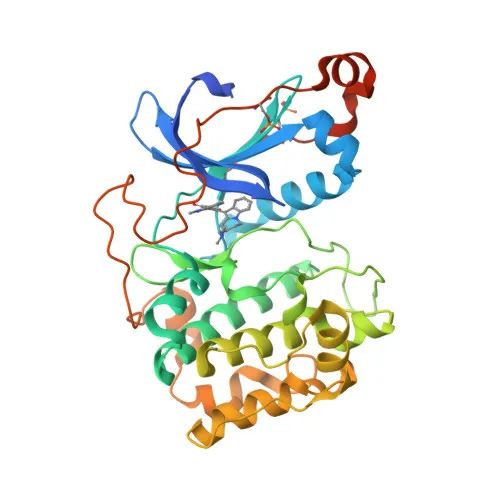
Phosphorylated Antibodies
Phospho-PRKCQ-S676 Rabbit pAb - Q04759
Item Number : CM0001811
Price varies based on specs and customizations
- Application
- WB, ELISA
- Cross Reactivity
- Human
- Protein Weight
- 82kDa

Shipping:
Contact us to get shipping details Enjoy On-time Dispatch Guarantee.
Why Choose Us
Easy ordering process, quality products, and dedicated support for your business success.
Core Product Specifications and Parameters
| Parameter | Value |
|---|---|
| Product Name | Phospho-PRKCQ-S676 Rabbit pAb |
| Remarks/Alias | PRKCT; nPKC-theta; Phospho-PRKCQ-S676 |
| Species | Human |
| Gene ID (Human) | 5588 |
| Gene ID | 5588 |
| Immunogen | Synthetic peptide corresponding to residues surrounding S676 of human PRKCQ (phospho-specific) |
| Source | Rabbit |
| Category | Phosphorylated Antibodies |
| Application | WB, ELISA |
| Cross Reactivity | Human |
| SWISS | Q04759 |
| Protein Weight | 82kDa |
| Shipping | Ice bag |
Biological Background: PRKCQ Function and Localization
- PRKCQ (Protein kinase C theta type), also known as nPKC-theta, is a calcium-independent, phospholipid- and diacylglycerol-dependent serine/threonine-protein kinase encoded by the PRKCQ gene. It plays non-redundant roles in T-cell receptor (TCR) signaling, essential for T-cell activation, proliferation, differentiation, and survival. Related references: PMID:21964608
- In TCR-CD3/CD28 co-stimulated T-cells, PRKCQ is required for the activation of NF-kappa-B and JUN, leading to IL2 production, and participates in calcium-dependent NFATC1 and NFATC2 transactivation. Related references: PMID:21964608
- PRKCQ directly phosphorylates CARD11 on serine residues, promoting CARD11 association with lipid rafts and recruitment of the BCL10-MALT1 complex to activate the canonical NF-kappa-B pathway. It also phosphorylates STK39/SPAK to mediate JUN activation. Related references: PMID:21964608
- Beyond T-cells, PRKCQ regulates signal transduction in platelets downstream of ITGA2B, CD36/GP4, F2R/PAR1, and F2RL3/PAR4 receptors, playing a positive role in 'outside-in' signaling and granule secretion. It may also inhibit insulin signaling by phosphorylating IRS1. Additionally, PRKCQ phosphorylates MSN, PDPK1, CCDC88A/GIV, and LRRK1, impacting various cellular processes. Related references: PMID:23509302, PMID:36040231
- Subcellular localization is predominantly cytoplasmic, with stimulus-dependent translocation to the cell membrane, consistent with its role in transducing signals from cell surface receptors.
- Tissue expression is prominent in skeletal muscle, T-cells, megakaryoblastic cells, and platelets, aligning with its key functions in immune cells and muscle.
- This antibody is raised against a phospho-peptide corresponding to residues surrounding serine 676 of human PRKCQ, enabling specific detection of the phosphorylated form. Serine 676 is a critical phosphorylation site involved in PRKCQ activation and downstream signaling.
- Post-translational modifications of PRKCQ include phosphorylation, ubiquitination, and isopeptide bond formation, as indicated by the keywords Phosphoprotein, Isopeptide bond, and Ubl conjugation. The protein also features zinc-finger domains, ATP-binding, and kinase activity.
Experimental Guidance and Technical Tips
- For Western blot applications, include phosphatase inhibitors in lysis buffers to preserve the phosphorylation status of PRKCQ at Ser-676.
- The phospho-specific nature of the antibody requires careful validation; consider using a non-phospho peptide blocking control or phosphatase treatment to confirm specificity.
- In ELISA, coating with phosphorylated versus non-phosphorylated peptides can help assess antibody selectivity.
- As a rabbit polyclonal antibody, detection requires an anti-rabbit IgG secondary conjugate; optimize primary and secondary antibody dilutions empirically for each assay.
- Given the 82 kDa molecular weight of PRKCQ, choose appropriate gel percentages and transfer conditions to ensure efficient resolution and detection.
CamelBio: Your One-Stop Sourcing Bridge
CamelBio serves as a comprehensive sourcing partner for IVD raw materials, offering validated antibodies, bulk reagents, and tailored procurement services. This phospho-specific PRKCQ antibody supports research and diagnostic workflows in T-cell signaling and inflammation. CamelBio can assist with scalable supply, custom antibody development, and technical consultation to accelerate your assay development from concept to clinic.
REQUEST A QUOTE
Our professional team will reply to you within one business day. Please feel free to contact us!