Phosphorylated Antibodies
Phospho-MAP2K1-S217/MAP2K2-S221 Rabbit Polyclonal Antibody for WB, ELISA - Q02750 / P36507
Item Number : CM0001753
Price varies based on specs and customizations
- Application
- WB, ELISA
- Cross Reactivity
- Human, Mouse, Rat
- Protein Weight
- 40kDa/43kDa/44kDa

Shipping:
Contact us to get shipping details Enjoy On-time Dispatch Guarantee.
Why Choose Us
Easy ordering process, quality products, and dedicated support for your business success.
Core Product Specifications and Parameters
| Parameter | Value |
|---|---|
| Product Name | Phospho-MAP2K1-S217/MAP2K2-S221 Rabbit pAb |
| Remarks/Alias | MEK1/MEK2; Phospho-MAP2K1-S217/MAP2K2-S221 |
| Species | Human |
| Gene ID (Human) | 5604/5605 |
| Immunogen | A synthetic phosphorylated peptide around S217 & S221 of human MAP2K1/MAP2K2 |
| Source | Rabbit |
| Category | Phosphorylated Antibodies |
| Application | WB, ELISA |
| Cross Reactivity | Human, Mouse, Rat |
| SWISS | Q02750/P36507 |
| Protein Weight | 40kDa/43kDa/44kDa |
| Shipping | Ice bag |
Biological Background: MAP2K1/MAP2K2 (MEK1/MEK2) Function and Localization
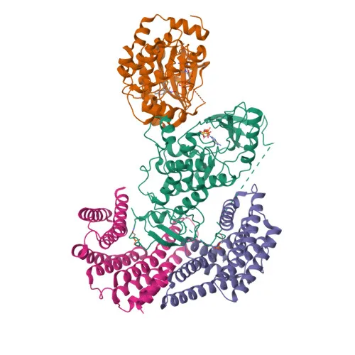
- MAP2K1 (MEK1) and MAP2K2 (MEK2) are dual-specificity protein kinases that serve as essential components of the MAP kinase signal transduction pathway, specifically the MAPK/ERK cascade.
- Upon extracellular stimulation, RAS activates RAF1, which in turn phosphorylates and activates MAP2K1/2 at conserved serine residues (S217 in MEK1, S221 in MEK2), triggering the downstream phosphorylation of ERK1/2 on a Thr-Glu-Tyr motif.
- Activated ERK1/2 then translocates to the nucleus to regulate transcription factors, controlling processes such as cell growth, adhesion, survival, differentiation, metabolism, and cytoskeletal rearrangements.
- MAP2K1 also participates in KSR1/2-dependent BRAF activation by promoting KSR-BRAF dimerization, which releases inhibitory interactions and enhances BRAF kinase activity. Related references: PMID:29433126
- The MAPK/ERK cascade influences endosomal dynamics, lysosome processing, and Golgi fragmentation during mitosis, mediated in part by MAP2K1's subcellular localization to the cytoplasm, centrosome, spindle pole, nucleus, and membrane.
- MAP2K1 is widely expressed across human tissues but exhibits extremely low levels in the brain, consistent with its ubiquitous role in mitogenic signaling.
- Key post-translational modifications include phosphorylation at activating sites, acetylation, and disease-associated mutations linked to cardiomyopathy, ectodermal dysplasia, and intellectual disability (per UniProt keywords).
Experimental Guidance and Technical Tips
- The immunogen is a synthetic phosphorylated peptide spanning S217 of MAP2K1 and S221 of MAP2K2, suggesting the antibody preferentially recognizes the activated (phosphorylated) forms of MEK1/2.
- For Western blot, prepare lysates from serum-starved cells stimulated with growth factors (e.g., EGF, insulin) to induce robust MEK1/2 phosphorylation; validate specificity by comparing with total MEK1/2 antibodies or treating samples with lambda phosphatase.
- Expected molecular weights are ~40–44 kDa for MAP2K1 and ~45 kDa for MAP2K2; alternative splicing may yield additional bands.
- In ELISA, the antibody can be used as a capture or detection reagent, but optimal dilution and pairing should be determined empirically in the final assay system.
CamelBio: Your One-Stop Sourcing Bridge
CamelBio is a dedicated one-stop sourcing partner for IVD raw materials, offering diagnostic manufacturers and researchers seamless access to validated antibodies, antigen pairs, and ancillary reagents from concept to clinic. This anti-phospho-MEK1/2 antibody is well-suited for studying MAPK/ERK pathway activation in cancer, development, and other signaling research, and CamelBio can facilitate bulk supply, custom conjugation, and rare-target reagent sourcing to support your assay development pipeline.
Product Datasheet
Phospho-MAP2K1-S217/MAP2K2-S221 Rabbit Polyclonal Antibody for WB, ELISA - Q02750 / P36507
REQUEST A QUOTE
Our professional team will reply to you within one business day. Please feel free to contact us!