Phosphorylated Antibodies
Phospho-IκBα-S36 Rabbit Monoclonal Antibody for WB, IHC-P, ELISA - P25963
Item Number : CM0001520
Price varies based on specs and customizations
- Application
- WB, IHC-P, ELISA
- Cross Reactivity
- Human, Mouse, Rat
- Protein Weight
- 36kDa

Shipping:
Contact us to get shipping details Enjoy On-time Dispatch Guarantee.
Why Choose Us
Easy ordering process, quality products, and dedicated support for your business success.
Core Product Specifications and Parameters
| Parameter | Value |
|---|---|
| Product Name | Phospho-IκBα-S36 Rabbit mAb |
| Remarks/Alias | IKBA; MAD-3; NFKBI; EDAID2; Phospho-IκBα-S36 |
| Species | Human |
| Gene ID (Human) | 4792 |
| Gene ID | 4792 |
| Immunogen | Synthetic Peptide: A synthetic phosphorylated peptide around S36 of human IκBα (P25963). |
| Source | Rabbit |
| Category | Phosphorylated Antibodies |
| Application | WB, IHC-P, ELISA |
| Cross Reactivity | Human, Mouse, Rat |
| SWISS | P25963 |
| Protein Weight | 36kDa |
| Shipping | Ice bag |
Biological Background: IκBα Function and Localization
-
IκBα (NF-kappa-B inhibitor alpha, also known as MAD3 or NFKBI) is a key regulatory protein that inhibits NF-κB transcription factors by retaining RELA/p65 and NFKB1/p50 dimers in the cytoplasm through masking their nuclear localization signals. Related references: PMID:1493333 PMID:36651806 PMID:7479976
-
Upon cellular stimulation by immune and pro-inflammatory signals, IκBα undergoes phosphorylation at Ser32 and Ser36, which triggers its ubiquitination and subsequent proteasomal degradation, thereby releasing NF-κB dimers to translocate to the nucleus and activate transcription. Related references: PMID:7479976 PMID:7628694 PMID:7796813
-
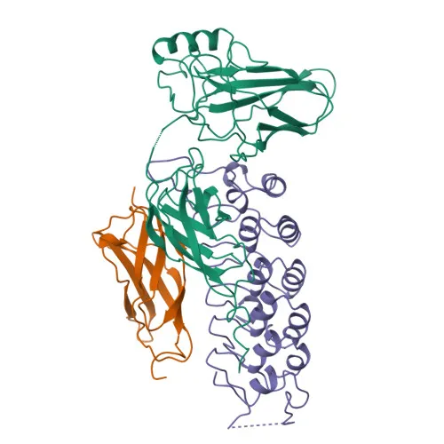
IκBα is a member of the IκB family of inhibitors, characterized by ankyrin repeats that mediate protein-protein interactions with NF-κB dimers.
-
Subcellularly, IκBα shuttles between the cytoplasm and the nucleus, but its predominant localization is cytoplasmic where it sequesters inactive NF-κB complexes.
-
Post-translational modifications of IκBα include phosphorylation, ubiquitination (Ubl conjugation), hydroxylation, and isopeptide bond formation, which regulate its stability and activity.
-
The gene NFKBIA is associated with ectodermal dysplasia and other immune-related disorders when mutated, highlighting its critical role in immune homeostasis.
Experimental Guidance and Technical Tips
- The immunogen is a synthetic phosphorylated peptide around Ser36, ensuring high specificity for the phosphorylated form. Non-phospho peptide blocking may be used to confirm phospho-specificity in WB.
- For WB, consider stimulating cells (e.g., with TNF-α or IL-1β) to induce IκBα phosphorylation and degradation, which can be monitored with this antibody.
- In IHC-P, antigen retrieval and permeabilization protocols should be optimized; validate staining in tissue sections known to express activated NF-κB signaling.
- For ELISA, use this antibody as a capture or detection reagent; cross-reactivity with unphosphorylated IκBα should be assessed depending on assay format.
- Because the antibody reacts with human, mouse, and rat, it is suitable for translational studies across these species; always include appropriate positive and negative controls.
CamelBio: Your One-Stop Sourcing Bridge
CamelBio serves as your integrated supply partner for IVD raw materials, offering ready-to-ship monoclonal antibodies, optimized antibody pairs, and ancillary reagents for NF-κB pathway research and diagnostic development. For this Phospho-IκBα-S36 antibody and related signaling targets, we can also source bulk quantities, rare specificities, and customized formats to streamline your assay pipeline from concept to clinic.
REQUEST A QUOTE
Our professional team will reply to you within one business day. Please feel free to contact us!