Phosphorylated Antibodies
Phospho-ERK1-T202 + ERK2-T185 Rabbit Polyclonal Antibody for WB, IHC-P, IF/ICC, ELISA - P28482 / P27361
Item Number : CM0001598
Price varies based on specs and customizations
- Application
- WB, IHC-P, IF/ICC, ELISA
- Cross Reactivity
- Human, Mouse, Rat
- Protein Weight
- 38kDa/40kDa/43kDa

Shipping:
Contact us to get shipping details Enjoy On-time Dispatch Guarantee.
Why Choose Us
Easy ordering process, quality products, and dedicated support for your business success.
Core Product Specifications and Parameters
| Parameter | Value |
|---|---|
| Product Name | Phospho-ERK1-T202 + ERK2-T185 Rabbit pAb |
| Species | Human |
| Gene ID | 5594 / 5595 |
| Immunogen | Synthetic phosphorylated peptide around T202 of human ERK1/ERK2 (NP_002737.2) |
| Source | Rabbit |
| Category | Phosphorylated Antibodies |
| Application | WB, IHC-P, IF/ICC, ELISA |
| Cross Reactivity | Human, Mouse, Rat |
| SWISS | P28482 / P27361 |
| Protein Weight | 38kDa/40kDa/43kDa |
| Shipping | Ice bag |
Biological Background: ERK1/ERK2 Function and Localization
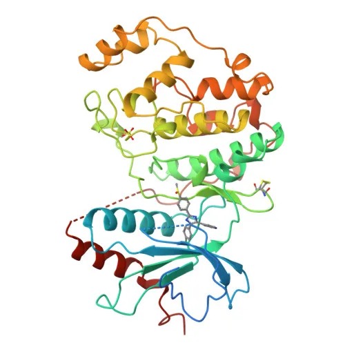
- ERK2 (MAPK1, also known as p42-MAPK) and ERK1 (MAPK3, p44-MAPK) are the two principal mitogen-activated protein kinases that constitute the core of the MAPK/ERK signaling cascade, transducing extracellular signals to regulate cell fate.
- The MAPK/ERK pathway controls a wide spectrum of cellular processes including proliferation, differentiation, adhesion, survival, and apoptosis by phosphorylating over 160 downstream substrates, such as transcription factors (e.g., ELK1, FOS, ATF2), protein kinases (e.g., RSK, MSK, MNK), and cytoskeletal regulators.
- Upon activation, ERK1/2 phosphorylates GLI1 to promote its dissociation from SUFU and nuclear translocation, thereby positively modulating smoothened signaling.
Related references: PMID:35831023 - In response to hypoxia, ERK2 phosphorylates phosphoglycerate kinase PGK1, directing it to the mitochondrion to suppress acetyl-CoA production from pyruvate.
Related references: PMID:26942675 - The kinase localizes to multiple subcellular compartments: cytoplasm, nucleus, cytoskeleton (spindle, centrosome), focal adhesions, and membrane caveolae, enabling context‑dependent signaling.
- Activation requires dual phosphorylation at the Thr-Glu-Tyr motif (T185 and Y187 in ERK2; T202 and Y204 in ERK1) by upstream MEK kinases; this antibody specifically detects the phosphorylated T185 (ERK2) and T202 (ERK1).
- Beyond kinase activity, ERK2 also functions as a transcriptional repressor by binding GC-rich promoter elements independently of its catalytic activity, modulating interferon‑responsive genes.
- Keywords: Acetylation, Alternative splicing, Apoptosis, ATP-binding, Cell cycle, Cell junction, Cytoplasm, Cytoskeleton, DNA-binding, Host-virus interaction, Kinase, Membrane, Nucleotide-binding, Nucleus, Phosphoprotein, Repressor, Serine/threonine-protein kinase, Transcription, Transcription regulation, Ubl conjugation.
Experimental Guidance and Technical Tips
- To preserve phosphorylation during sample preparation, include phosphatase inhibitors in lysis buffers and avoid prolonged sample handling at room temperature.
- Validate antibody specificity by treating cell lysates with lambda protein phosphatase; expect complete loss of signal in Western blot.
- In IHC-P, optimize antigen retrieval conditions (e.g., citrate buffer pH 6.0) and consider using a phospho‑specific signal amplification protocol to enhance detection of low‑abundance phosphorylated targets.
- For IF/ICC, fix cells with formaldehyde and permeabilize with methanol; confirm colocalization with known subcellular markers (e.g., nuclear DAPI, Golgi markers) based on the expected localization upon stimulation.
- Expected bands: ~38–43 kDa for ERK1/ERK2 isoforms; multiple bands may appear due to phosphorylation states or splice variants.
CamelBio: Your One-Stop Sourcing Bridge
CamelBio provides diagnostic manufacturers and research laboratories with reliable access to high‑quality IVD raw materials, including this phospho‑specific ERK1/ERK2 antibody. Our portfolio covers validated antibody pairs, recombinant antigens, and ancillary reagents to support MAPK signaling‑related assay development. With one‑stop sourcing and consultative technical support, CamelBio helps you translate basic research into robust diagnostic solutions.
REQUEST A QUOTE
Our professional team will reply to you within one business day. Please feel free to contact us!