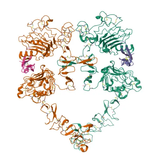
Phosphorylated Antibodies
Phospho-EGFR-Y869 Rabbit Polyclonal Antibody for WB and ELISA - P00533
Item Number : CM0001803
Price varies based on specs and customizations
- Application
- WB, ELISA
- Cross Reactivity
- Human, Mouse, Rat
- Protein Weight
- 134kDa

Shipping:
Contact us to get shipping details Enjoy On-time Dispatch Guarantee.
Why Choose Us
Easy ordering process, quality products, and dedicated support for your business success.
Core Product Specifications and Parameters
| Parameter | Value |
|---|---|
| Product Name | Phospho-EGFR-Y869 Rabbit pAb |
| Remarks/Alias | ERBB; ERRP; HER1; mENA; ERBB1; PIG61; NISBD2; Phospho-EGFR-Y869 |
| Species | Human |
| GeneID (Human) | 1956 |
| GeneID | 1956 |
| Immunogen | Synthetic Peptide: A synthetic phosphorylated peptide around Y869 of human EGFR (NP_005219.2) |
| Source | Rabbit |
| Category | Phosphorylated Antibodies |
| Application | WB, ELISA |
| Cross Reactivity | Human, Mouse, Rat |
| SWISS | P00533 |
| Protein Weight | 134kDa |
| Shipping | Ice bag |
Biological Background: EGFR Function and Localization
-
EGFR is a receptor tyrosine kinase that binds EGF family ligands (including EGF, TGF-alpha, AREG, epigen, betacellulin, epiregulin, and HBEGF), leading to receptor dimerization and autophosphorylation. Related references: PMID:10805725 PMID:27153536 PMID:2790960 PMID:12297049 PMID:15611079 PMID:17909029
-
Ligand-activated EGFR triggers multiple intracellular signaling cascades, including the RAS-RAF-MEK-ERK, PI3 kinase-AKT, PLCgamma-PKC, and STAT modules, as well as NF-kappa-B signaling. Related references: PMID:27153536 PMID:11116146
-
EGFR directly phosphorylates other proteins such as RGS16 (enhancing its GTPase activity) and MUC1 (promoting its interaction with SRC and beta-catenin), linking to G protein and cell adhesion pathways. Related references: PMID:11602604 PMID:11483589
-
EGFR positively regulates cell migration by interacting with CCDC88A/GIV, which retains EGFR at the cell membrane and sustains signaling. Related references: PMID:20462955
-
The EGFR gene (also known as ERBB1, HER1) encodes a receptor tyrosine kinase that belongs to the ERBB family; an alternatively spliced isoform (isoform 2) may act as an antagonist of EGF action and is expressed in ovarian cancers.
-
EGFR is predominantly localized at the cell membrane but is also detected in the endoplasmic reticulum, Golgi apparatus, nucleus membrane, endosomes, and can be secreted.
-
EGFR is a phosphoprotein with key tyrosine phosphorylation sites (including Y869) that regulate its kinase activity, and undergoes glycosylation, ubiquitination, and other post-translational modifications.
-
Ubiquitously expressed in human tissues; EGFR acts as a host cell receptor for hepatitis C virus (HCV), facilitating viral entry into hepatocytes.
Experimental Guidance and Technical Tips
- The immunogen is a synthetic phosphopeptide corresponding to residues around Y869 of human EGFR. To confirm phospho-specificity, include phosphatase-treated controls or compete with non-phosphorylated peptide.
- For western blot, use EGF-stimulated cell lysates and expect a band at ~134 kDa; cross-reactivity with mouse and rat EGFR is reported, but validate in your species of interest.
- For ELISA, consider coating with a phosphorylated peptide and optimizing blocking and detection conditions to enhance signal-to-noise ratio.
- Avoid harsh stripping conditions when reprobing blots, as this may remove the phospho-specific signal.
CamelBio: Your One-Stop Sourcing Bridge
CamelBio offers a comprehensive range of IVD raw materials, including validated phospho-specific antibodies, recombinant proteins, and ancillary reagents. For EGFR signaling research and diagnostic assay development, we provide reliable, high-quality antibodies and can support sourcing for rare or custom targets. From initial concept to clinical application, CamelBio streamlines your supply chain as a trusted partner.
REQUEST A QUOTE
Our professional team will reply to you within one business day. Please feel free to contact us!