Phosphorylated Antibodies
Phospho-CDK1-T161+CDK2/CDK3-T160 Rabbit Monoclonal Antibody for WB, IP, IF - P06493
Item Number : CM0000667
Price varies based on specs and customizations
- Application
- WB, IP, IF-P, IHC-P, DB, ELISA
- Cross Reactivity
- Human, Mouse, Rat
- Protein Weight
- 34kDa

Shipping:
Contact us to get shipping details Enjoy On-time Dispatch Guarantee.
Why Choose Us
Easy ordering process, quality products, and dedicated support for your business success.
Core Product Specifications and Parameters
| Parameter | Value |
|---|---|
| Product Name | Phospho-CDK1-T161+CDK2/CDK3-T160 Rabbit mAb |
| Remarks/Alias | CDC2; CDC28A; P34CDC2; CDK1 |
| Species | Human |
| GeneID (Human) | 983 |
| GeneID | 983 |
| Immunogen | Synthetic peptide: A synthetic phosphorylated peptide around T161 of human CDK1 (NP_001777.1). |
| Source | Rabbit |
| Category | Phosphorylated Antibodies |
| Application | WB, IP, IF-P, IHC-P, DB, ELISA |
| Cross Reactivity | Human, Mouse, Rat |
| SWISS | P06493 |
| Protein Weight | 34kDa |
| Shipping | Ice bag |
Biological Background: CDK1 Function and Localization
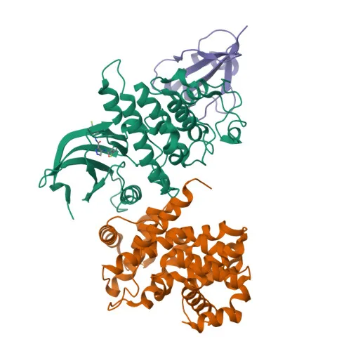
- Cyclin-dependent kinase 1 (CDK1, also known as CDC2/p34) is a highly conserved serine/threonine kinase that serves as the master regulator of the eukaryotic cell cycle, driving the G2-M transition and coordinating mitotic entry and progression (PubMed:16407259, PubMed:16933150, PubMed:17459720). Related references: PMID:16407259 PMID:16933150 PMID:17459720
- CDK1 activity is tightly controlled by cyclin binding and phosphorylation: activation requires T161 phosphorylation by the CDK-activating kinase (CAK) complex (CDK7/cyclin H/MAT1), while inhibitory phosphorylation at T14 and Y15 is mediated by WEE1 and PKMYT1; dephosphorylation by CDC25 phosphatases at the G2-M boundary triggers sudden activation (PubMed:18480403, PubMed:20360007, PubMed:2188730). Related references: PMID:18480403 PMID:20360007 PMID:2188730
- Once activated, CDK1 phosphorylates over a hundred downstream substrates to orchestrate mitotic events, including lamin phosphorylation leading to nuclear envelope breakdown, microtubule-associated protein phosphorylation for spindle assembly, and histone H1 phosphorylation for chromosome condensation (PubMed:2188730, PubMed:2344612, PubMed:37788673). Related references: PMID:2188730 PMID:2344612 PMID:37788673
- Subcellularly, CDK1 dynamically localizes to the nucleus, cytoplasm, mitochondria, centrosomes, and mitotic spindle, reflecting its diverse functional roles during cell cycle progression.
- CDK1 also regulates transcription factors (e.g., FOXO1), apoptotic proteins (e.g., Bcl-xL, caspase 8), and epigenetic modifiers (e.g., EZH2), linking cell cycle control to cell fate decisions and DNA damage responses (PubMed:18356527, PubMed:19917720, PubMed:20937773, PubMed:20935635). Related references: PMID:18356527 PMID:19917720 PMID:20937773 PMID:20935635
- CDK1 is essential for early embryonic development and its dysregulation is frequently observed in cancer, where it may be overexpressed or hyperactivated, contributing to uncontrolled proliferation (PubMed:18480403, PubMed:20360007). Related references: PMID:18480403 PMID:20360007
- Additionally, CDK1 serves as a receptor for hepatitis C virus (HCV) entry into hepatocytes, suggesting a role beyond cell cycle regulation.
Experimental Guidance and Technical Tips
- The immunogen is a synthetic phosphopeptide targeting phosphorylated T161 of human CDK1, a conserved residue in the activation loop. The antibody is expected to recognize the activated (T161-phosphorylated) form of CDK1 and potentially cross-react with phosphorylated T160 of CDK2 and CDK3 due to sequence homology.
- For Western blotting, treat cell lysates with phosphatase inhibitors to preserve phosphorylation status. The predicted molecular weight is ~34 kDa; monitor band shifts or intensity changes upon phosphatase treatment to confirm phospho-specificity.
- In immunoprecipitation, pre-clearing with a control IgG and using non-denaturing lysis conditions may improve detection of CDK1 complexes.
- For immunofluorescence and immunohistochemistry, permeabilization and fixation methods should be optimized; the antibody may detect nuclear, cytoplasmic, and centrosomal staining patterns depending on cell cycle phase.
- Dot blot and ELISA applications can be used for high-throughput screening of phospho-CDK1 levels in samples.
CamelBio: Your One-Stop Sourcing Bridge
CamelBio is dedicated to providing diagnostic manufacturers, laboratories, and research institutes with a one-stop supply of IVD raw materials, technical services, and consulting support from concept to clinic. Our portfolio includes validated antibody pairs, optimized monoclonal and polyclonal antibodies, bulk ancillary reagents, and custom rare target sourcing. This anti-phospho-CDK1 antibody supports cell cycle and oncology research applications, and we offer flexible procurement solutions to accelerate your development and manufacturing workflows.
Product Datasheet
Phospho-CDK1-T161+CDK2/CDK3-T160 Rabbit Monoclonal Antibody for WB, IP, IF - P06493
REQUEST A QUOTE
Our professional team will reply to you within one business day. Please feel free to contact us!