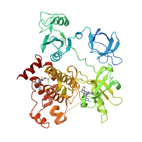
Phosphorylated Antibodies
Phospho-c-Abl-Y412 Rabbit pAb - P00519
Item Number : CM0000491
Price varies based on specs and customizations
- Application
- IHC-P, ELISA
- Cross Reactivity
- Human, Mouse, Rat
- Protein Weight
- 123kDa

Shipping:
Contact us to get shipping details Enjoy On-time Dispatch Guarantee.
Why Choose Us
Easy ordering process, quality products, and dedicated support for your business success.
Core Product Specifications and Parameters
| Parameter | Value |
|---|---|
| Product Name | Phospho-c-Abl-Y412 Rabbit pAb |
| Remarks/Alias | ABL; JTK7; p150; c-ABL; v-abl; CHDSKM; c-ABL1; BCR-ABL; bcr/abl; Phospho-c-Abl-Y412 |
| Species | Human |
| GeneID (human) | 25 |
| GeneID | 25 |
| Immunogen | Synthetic peptide|A synthetic phosphorylated peptide around Y412 of human ABL1 (NP_009297.2) |
| Source | Rabbit |
| Category | Phosphorylated Antibodies |
| Application | IHC-P, ELISA |
| Cross Reactivity | Human, Mouse, Rat |
| SWISS | P00519 |
| Protein Weight | 123kDa |
| Shipping | Ice bag |
Biological Background: c-Abl Function and Localization
- c-Abl, encoded by ABL1, is a non-receptor tyrosine-protein kinase proto-oncogene also known as Abelson tyrosine-protein kinase 1, p150, and BCR-ABL (in leukemogenic fusions).
- It coordinates actin cytoskeleton remodeling, cell motility, adhesion, and endocytosis by phosphorylating effectors such as WASF3, DBNL, CTTN, and ENAH (PubMed:22810897). Related references: PMID:22810897
- In response to oxidative stress, ABL1 translocates to mitochondria, phosphorylates PRKD2 on Tyr‑717, and promotes mitochondrial dysfunction and apoptosis (PubMed:28428613). Related references: PMID:28428613
- Nuclear ABL1 participates in DNA‑damage sensing and repair, interacting with factors such as DDB1, RAD51, and WRN; it also triggers TP73‑dependent apoptosis when damage is overwhelming.
- A critical regulatory autophosphorylation site is Tyr‑412 (the target of this antibody), which lies in the activation loop and is essential for kinase activity.
- ABL1 is widely expressed and localizes to the cytoplasm, cytoskeleton, nucleus, and mitochondria, reflecting its diverse roles.
- It carries numerous post‑translational modifications (phosphorylation, acetylation, myristoylation, ubiquitination) and interacts with SH2/SH3‑domain‑containing adaptor proteins.
- Pathogens such as Helicobacter pylori (CagA) and Vaccinia virus hijack ABL1 signaling to reorganize the host actin cytoskeleton for infection.
Experimental Guidance and Technical Tips
- This antibody was raised against a synthetic phosphopeptide corresponding to Tyr‑412 of human ABL1; we recommend validating specificity by comparing signals in untreated vs. phosphatase‑treated lysates.
- For IHC‑P, consider standard antigen retrieval methods (e.g., citrate buffer, pH 6.0) and test a range of primary antibody dilutions to optimize phospho‑signal detection.
- In ELISA, use phosphorylated and non‑phosphorylated peptide controls to confirm phospho‑specificity and cross‑reactivity.
- The reported cross‑reactivity with mouse and rat suggests conserved epitope sequences; however, always verify reactivity in your specific model system.
CamelBio: Your One-Stop Sourcing Bridge
CamelBio provides diagnostic and life science manufacturers streamlined access to critical IVD raw materials, including validated phospho‑specific antibodies like this Phospho‑c‑Abl‑Y412 Rabbit pAb. Our platform supports your complete workflow—from optimized monoclonal and polyclonal antibodies to bulk ancillary reagents and custom sourcing of rare‑target proteins. Let CamelBio be your research and development partner, helping you bridge concept to clinic with confidence.
REQUEST A QUOTE
Our professional team will reply to you within one business day. Please feel free to contact us!