Phosphorylated Antibodies
Phospho-ATF2 (T69/T71) Rabbit pAb for WB, IHC-P, ELISA - P15336
Item Number : CM0000866
Price varies based on specs and customizations
- Application
- WB, IHC-P, ELISA
- Cross Reactivity
- Human, Mouse, Rat
- Protein Weight
- 55kDa

Shipping:
Contact us to get shipping details Enjoy On-time Dispatch Guarantee.
Why Choose Us
Easy ordering process, quality products, and dedicated support for your business success.
Core Product Specifications and Parameters
| Parameter | Value |
|---|---|
| Product Name | Phospho-ATF2-T69/T71 Rabbit pAb |
| Remarks/Alias | HB16; CREB2; TREB7; CREB-2; CRE-BP1; Phospho-ATF2-T69/T71 |
| Species | Human |
| GeneID (Human) | 1386 |
| GeneID | 1386 |
| Immunogen | A synthetic phosphorylated peptide around T69 & T71 of human ATF2 (NP_001871.2). |
| Source | Rabbit |
| Category | Phosphorylated Antibodies |
| Application | WB, IHC-P, ELISA |
| Cross Reactivity | Human, Mouse, Rat |
| SWISS | P15336 |
| Protein Weight | 55kDa |
| Shipping | Ice bag |
Biological Background: ATF2 Function and Localization
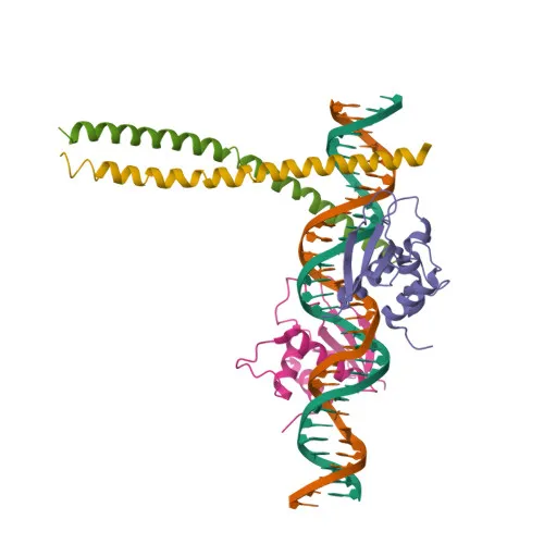
- Activating transcription factor 2 (ATF-2), also known as HB16, CREB2, TREB7, or CRE-BP1, is a member of the ATF/CREB family of transcription factors. The gene undergoes alternative splicing, yielding multiple isoforms.
- ATF-2 functions as a transcriptional activator by binding to CRE (cAMP response element) consensus sequences (5'-TGACGTCA-3') or AP-1 (activator protein 1) consensus sequences (5'-TGACTCA-3'), regulating genes involved in anti-apoptosis, cell growth, proliferation, and the DNA damage response.
- Subcellular localization includes the nucleus, cytoplasm, and mitochondrial outer membrane, enabling context-dependent functions in transcription, mitochondrial regulation, and apoptosis.
- In the cytoplasm, ATF-2 interacts with hexokinase-1 (HK1) and voltage-dependent anion channel 1 (VDAC1) at the mitochondrial outer membrane, disrupting mitochondrial membrane potential and promoting cell death.
- Phosphorylation at Thr69 and Thr71 by ATM kinase is critical for the DNA damage response. Phosphorylated ATF-2 participates in ionizing radiation (IR)-induced S phase checkpoint control and recruits the MRN complex to IR-induced foci.
- ATF-2 exhibits intrinsic histone acetyltransferase (HAT) activity, specifically acetylating histones H2B and H4 in vitro, linking transcription regulation to chromatin modification. Related references: PMID:10821277
- ATF-2 can display either oncogenic or tumor suppressor activities depending on tissue and cellular context. It is ubiquitously expressed, with relatively higher levels in the brain.
Experimental Guidance and Technical Tips
- For Western blot, consider using fresh protein samples with phosphatase inhibitors to preserve phosphorylation. Include a non-phosphorylated control to confirm phospho-specificity.
- In IHC-P, antigen retrieval methods may be required; titrate the antibody to minimize background, and validate using tissue known to express phospho-ATF2.
- For ELISA, coat plates with the phospho-peptide immunogen as a positive control, and validate cross-reactivity against the unphosphorylated peptide if needed.
- Given cross-reactivity with mouse and rat, verify sequence alignment of the immunogen region and validate performance in the relevant species and tissue models.
CamelBio: Your One-Stop Sourcing Bridge
CamelBio specializes in providing diagnostic manufacturers and research institutes with a comprehensive range of IVD raw materials, from validated antibody pairs to optimized monoclonal and polyclonal antibodies. This anti-phospho-ATF2 (T69/T71) antibody supports studies in transcription regulation, apoptosis, and DNA damage response. Beyond single reagents, we offer bulk ancillary reagents and tailored sourcing for rare targets, ensuring seamless support from concept to clinic.
REQUEST A QUOTE
Our professional team will reply to you within one business day. Please feel free to contact us!